Category: Science

  • [Science] A ‘DNA Typewriter’ Maps How One Cell Becomes a Mouse — Clues to Birth Defects, Cancer

    [Science] A ‘DNA Typewriter’ Maps How One Cell Becomes a Mouse — Clues to Birth Defects, Cancer

    Every mammal begins life as a single fertilized egg. Through repeated divisions and a long sequence of developmental decisions, that one cell gives rise to the many specialized cells that build an animal’s body. Reconstructing exactly how those cells are related to one another has been a major scientific challenge — researchers have never had a detailed, verified map of which cell gave rise to which inside a living mammal. Two research teams have now taken a significant step toward solving that problem, publishing independently in the same week. Using DNA-based molecular recorders built into mouse embryos, they worked backward from genetic marks left behind in cells to reconstruct how a single fertilized egg becomes a full body. The work could eventually help researchers investigate how birth defects, neurodevelopmental conditions, and cancer arise.

    A Lineage Record Written Into DNA

    The core tool, called DNA Typewriter, was developed by a University of Washington (UW) School of Medicine team that includes geneticist Jay Shendure and researcher Junhong Choi. It is built on prime editing, a precise form of CRISPR-based gene editing that rewrites short stretches of DNA without fully cutting both strands. The team engineered a short, designated sequence into cells’ genomes and set it up so that each cell division adds one new mark, in strict order, to that sequence. Because the marks accumulate like letters typed one after another, researchers can later read the resulting sequence in any surviving cell and reconstruct which cells share a recent common ancestor and which branched apart earlier — in effect, rebuilding a family tree after the fact, rather than filming it as it happens.

    Older lineage-tracing systems generally relied on CRISPR methods that cut DNA to create heritable barcodes. Those barcodes can be powerful, but as edits pile up they can become harder to read unambiguously, and some lineage information is inevitably lost along the way. Because DNA Typewriter writes new sequence without the same kind of DNA break, the team reports it can keep recording more steadily, at higher resolution, for longer than earlier cut-based approaches — though, like any method, its performance still depends on how efficiently it operates inside developing cells.

    One Richly Recorded Embryo

    In the new study, published October 8, 2026 in the journal Science, the UW-led team — including co-first authors Qi Yu, Haedong Kim and Sophie Seidel, with Chengxiang Qiu of Dartmouth College and Shendure as co-senior authors — attempted to activate DNA Typewriter recording in 100 fertilized mouse eggs. Only 10 developed into embryos that could later be examined, and of those, one — labeled Embryo No. 3 — carried by far the richest, most complete record. That embryo became the basis for the study’s detailed lineage reconstruction, which covered roughly 1.3 million individual embryonic cells.

    The recording spanned 13.5 days of development, from the fertilized egg’s first division through “late organogenesis,” the stage when the body plan and internal organs have largely taken shape. A full mouse pregnancy lasts 19 to 21 days, so the reconstruction covers a large share of gestation, though not the full life cycle. As co-first author Haedong Kim put it, the goal was a tool that “writes everything in strict order so that we can record cell lineages at much higher resolution for a longer time” than earlier barcoding methods allowed.
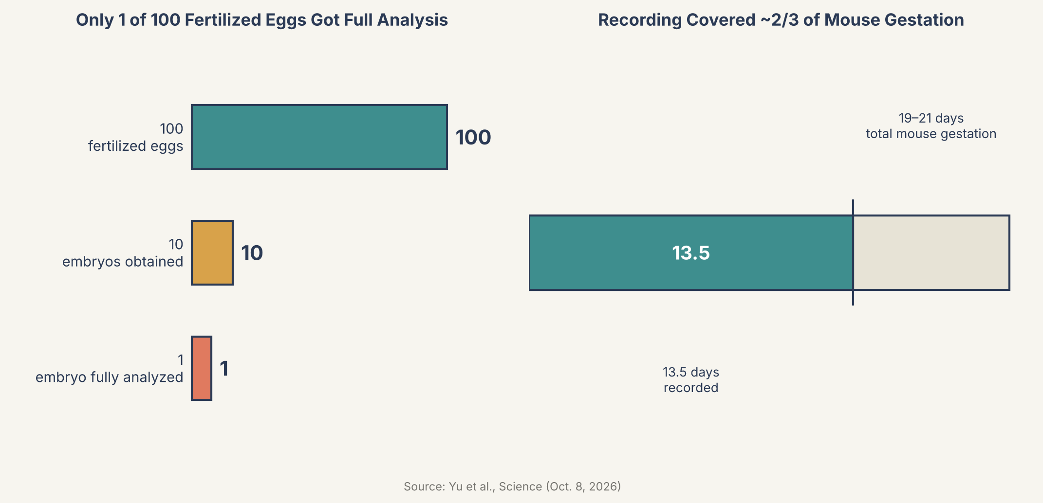
    Chart showing 100 fertilized eggs yielding 10 embryos and 1 fully analyzed, and 13.5 of 19-21 days of mouse gestation recorded
    Source: based on data from Yu et al., Science (Oct. 8, 2026)

    Two Founder Cells, One Shared Outcome

    A built-in mark distinguished the two cells created when the fertilized egg first divided, letting researchers trace nearly every profiled cell in the embryo back to one of those two founders. One founder went on to produce noticeably more descendant cells than the other. Yet both founders generated a broadly similar mix of cell types in roughly equal proportions — meaning that an early imbalance in how many cells each founder produced did not translate into one founder specializing in different tissue types than the other.

    The data also pointed to differences in timing: some blood and retinal cell populations showed signs of settling into a restricted identity relatively early in development, while cells contributing to the outer layer of skin appeared to keep their options open for longer. That kind of timing information — not just which cells are related, but roughly when their developmental paths narrowed — is difficult to extract from a single still snapshot of tissue, which is part of why a continuous lineage record is valuable.

    A Second, Larger-Scale Study Agrees

    The same week, a separate team led by Jonathan Weissman published a companion study in the journal Cell, using a related but distinct prime-editing-based system called PEtracer. Rather than following a single embryo in exhaustive detail, that study pooled data from 16 mouse embryos spanning days 7.5 to 10 of development, profiling more than 1.4 million cells in total to build a broader, population-level map of when and how cells commit to their fates. The two approaches are complementary: DNA Typewriter trades breadth for an unusually dense, ordered timeline in a single animal, while PEtracer trades timeline detail for scale across many animals. That two independently developed methods, from different labs, point toward a broadly consistent picture of mouse development adds useful weight to both — without, of course, each study individually confirming every specific claim made by the other.

    “This kind of large-scale developmental recording gives scientists a map of how one cell becomes a whole body,” says Haedong Kim, co-first author of the Science study.

    Weissman has framed the pair of studies as a long-sought proof of principle: that mammalian biology may finally be approaching something like the complete cell lineage map that biologist John Sulston built for the roundworm C. elegans back in 1983, by directly watching the worm’s roughly 1,000 cells divide under a microscope over years of painstaking observation. A mouse — and, by extension, a human — is built from many billions of cells, far beyond what direct visual tracking could ever handle. Genomic recording tools like DNA Typewriter and PEtracer offer a different route toward that same kind of detailed lineage map, built from molecular records and computational reconstruction rather than a person watching through a lens.
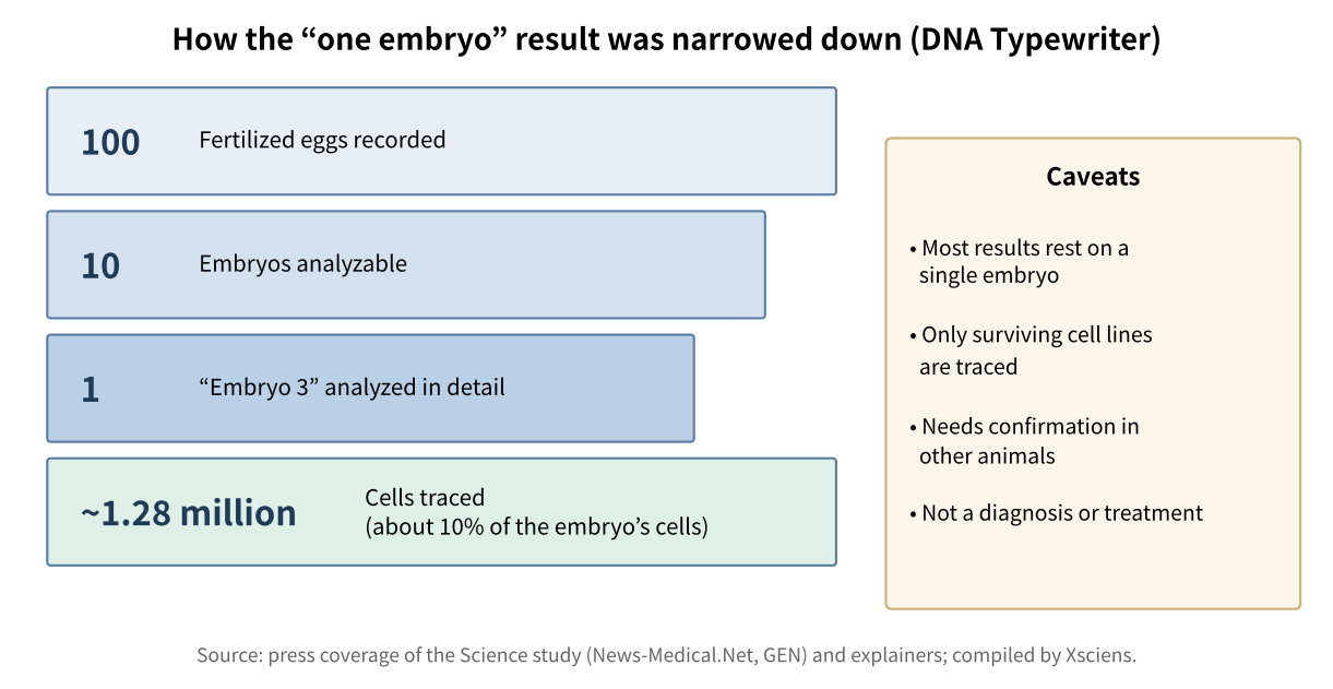
    Funnel from 100 fertilized eggs to 10 analyzable embryos, 1 detailed embryo and about 1.28 million traced cells, with caveats
    How the experiment was narrowed down, and how to read the result (compiled by Xsciens from press coverage and explainers).

    Why This Matters for Medicine

    The appeal of this kind of lineage map goes beyond curiosity about development for its own sake. If researchers can see roughly how and when healthy cells commit to becoming blood, skin, or retina, they gain a reference point for recognizing what might go differently in congenital malformations, in neurodevelopmental conditions linked to early brain development, and in genetic diseases more broadly — as a way to investigate how developmental abnormalities arise, not as a diagnosis or treatment in itself. Cancer researchers are watching this space too, since tumors can, in part, involve cells that lose normal control over their fate decisions, among other changes; fine-grained lineage data is one more tool for studying how abnormal cell populations evolve over time. Xsciens has previously reported on research into how an existing cancer drug affects brain tumor cells, a reminder that untangling cellular mechanisms in detail, lineage included, is an active front across cancer research generally. Longer term, scientists studying stem cells hope lineage maps like these could help guide how lab-grown cells are coaxed into becoming the right tissue for future cell-based therapies.

    It’s worth being clear about the limits here. The DNA Typewriter study’s richest results come from a single mouse embryo out of ten examined; broader validation across more animals, and eventually work beyond mice, will be needed before the tool becomes a routine research platform. Both systems have so far only been used in laboratory mouse embryos, not in humans, and translating developmental insights from mice to people remains its own multi-step scientific challenge. Even so, having two independent, broadly converging demonstrations published within the same week is a meaningful sign that genomic cell-lineage recording has moved from a promising idea toward a working research tool — one whose maps are highly informative, even if not yet complete.

    Xsciens Editorial Note

    “DNA wrote down how one egg becomes a body” is an appealing story, but it helps to weigh the numbers first.

    • What was shown. A method that writes marks into DNA without cutting it traced a “family tree” of about 1.28 million cells over 13.5 days. A separate team’s method reported similar results, so the direction has some support.
    • What is still open. The detailed analysis rests on one embryo out of 100 eggs, and the traced cells are about 10% of the embryo. Coverage of the study notes that dead cells leave no record and that confirmation in other individuals is needed.
    • A caution when reading. “Clues to birth defects and cancer” in the headline is about future uses. The study itself did not demonstrate any diagnosis or treatment.
    • Our imagination (speculation) — We wonder whether, as this family tree grows, disease researchers might be able to compare against a kind of standard map of “where does this cell come from when everything is normal?” That could make it easier to discuss at which branching step a congenital condition arises. But until the method works reliably across many individuals, that remains a long way off, and this is our speculation.

    References for this note: The Brighter Side, “Scientists construct a ‘family tree’ containing 1.28 million cells from a single embryo” (figures from the Science study and the limitations it lists)

    Sources: News-Medical.Net, Genetic Engineering & Biotechnology News (GEN)
  • AI Summaries Can Distort Human Memory — A Warning for Police and Witnesses

    AI Summaries Can Distort Human Memory — A Warning for Police and Witnesses

    You watch a short video of a car approaching an intersection, notice the traffic sign, and see it strike a pedestrian. The next day, you read an AI-generated summary of that video, one that happens to get the sign wrong. Asked afterward what you actually saw, you become measurably less likely to answer correctly. That is the core result of a new study from Georgetown University and the University of Washington: a misleading AI summary can distort people’s memory of an event they witnessed directly, which matters because AI-generated summaries are already spreading into newsrooms, offices, and policing.

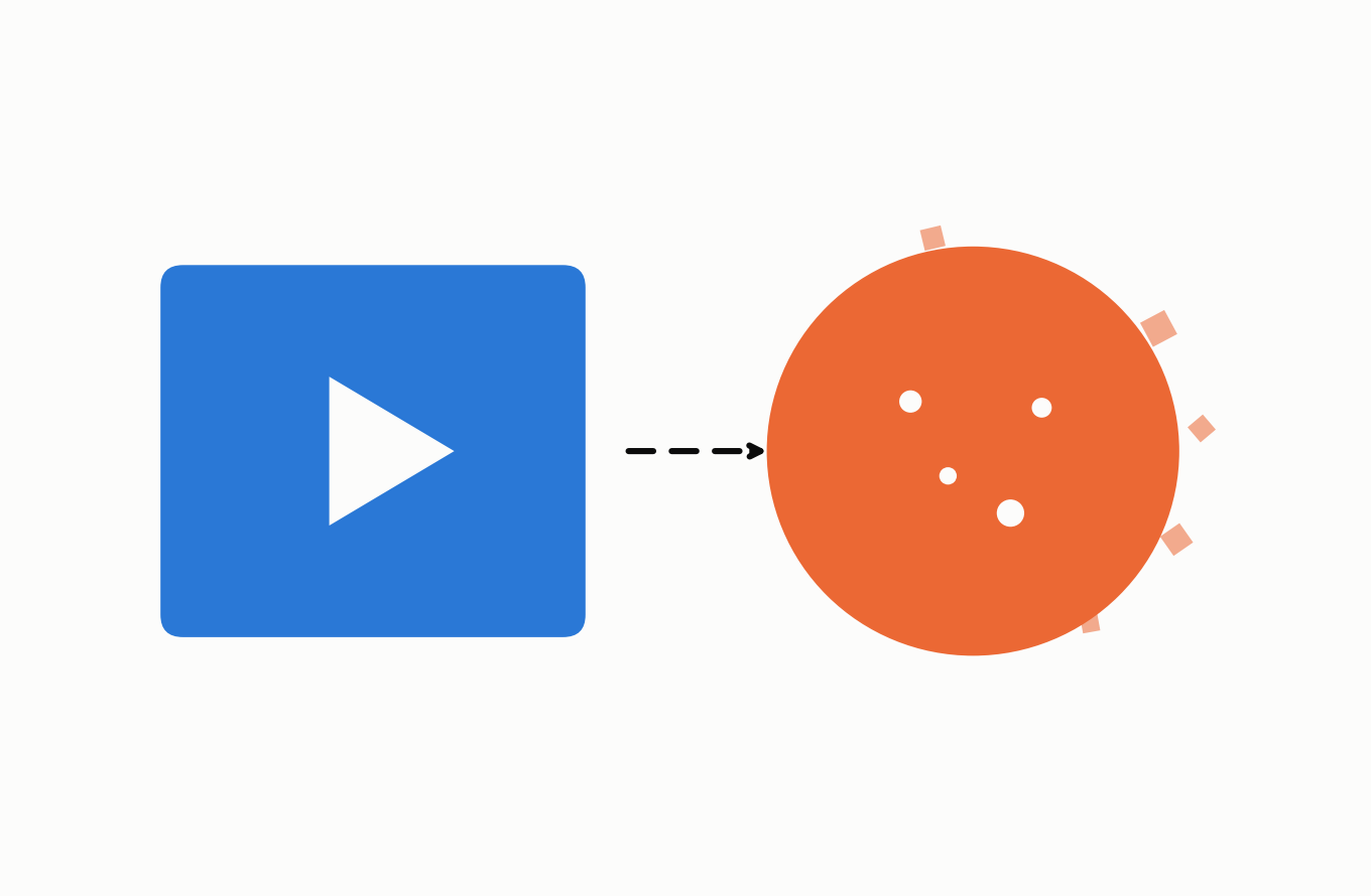
    Illustration: video summarized by AI, with details breaking off before reaching memory
    From video to AI summary to memory: small details can break off and go missing along the way — an illustration of the distortion effect described in this article.

    AI Summaries Are Already Everywhere

    Large language models like ChatGPT and Gemini are now routinely used to condense meeting recordings, news footage, and surveillance video into a few bullet points. Some U.S. police departments have gone further, adopting separate tools that draft incident-report narratives from transcribed body-camera audio. One such product, Axon’s Draft One, generates a draft within minutes of a call ending, which an officer must then review and approve; the Fort Collins, Colorado police department has reported an 82% drop in time spent writing reports after adopting it. That tool drafts text from audio transcripts rather than summarizing video, and it was not tested in this study — but it illustrates how quickly AI-written accounts are entering settings where accuracy carries real stakes.

    That broader shift is what worries the study’s authors. “AI is a new method of delivering misinformation,” said lead author Mattea Sim, an assistant research professor at Georgetown’s McCourt School of Public Policy. Co-author Yael Eiger, a Ph.D. candidate at the University of Washington who studies technology in policing, put it more bluntly: she said it concerns her that police departments may already be relying on video-summarization technology without the kind of testing this study attempted.

    Testing Memory With a Car-Crash Video

    The researchers ran their test in two stages. First, they asked ChatGPT-5.5 and Gemini 2.5 Flash-Lite to summarize two short animated videos of a car-pedestrian collision. The two videos were nearly identical except for one detail: one showed the car approaching a stop sign, the other a yield sign. Each model summarized each video five times, producing 20 summaries in total.

    Second, 360 people were recruited to watch one of the two videos themselves. One to two days later, the researchers had each participant read a summary of what they had just seen — either an accurate one or one with a single planted error that reversed the key sign detail — and told them, at random, that the summary had been written by either an AI system or a human transcriber. Of the 360 who started, 328 completed both stages and passed an attention check.

    Most Summaries Omitted the Key Event

    Before testing human memory, the researchers first graded the 20 AI-written summaries against a list of details the researchers had identified as central to the event. The results were not reassuring: on average, the summaries omitted 51.6% of those central details, and 19 of the 20 summaries failed to mention the single most important one — the car hitting the pedestrian.

    The two models differed in other ways. Every summary from both ChatGPT and Gemini contained at least one error. Gemini’s summaries included a critical inaccuracy — one that reversed a core fact, such as the traffic sign, rather than just dropping a minor detail — in all of its summaries, compared with 30% for ChatGPT. Both models also occasionally invented details that were never in the video at all, a pattern researchers call hallucination. These figures come from a small, controlled test of two models summarizing two short videos, so they should be read as evidence that serious errors can occur rather than as a general failure rate for AI video summaries.

    “AI has the potential to generate misinformation, even absent any adversarial intent,” the study authors write.

    A Wrong Summary Cut Recall Nearly in Half

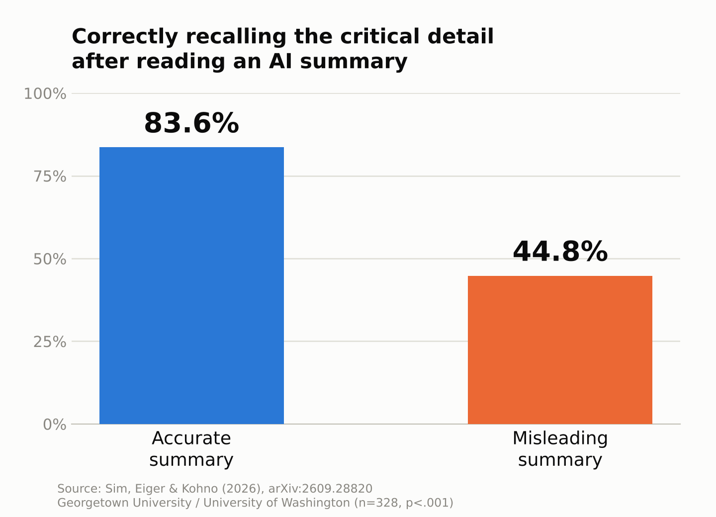
    The second stage showed these errors don’t stay confined to the summary — they appear to bleed into people’s own memory of the event. Participants who read an accurate summary correctly identified the sign 83.6% of the time. Among those who read the summary with the planted error, accuracy dropped to 44.8%, a gap the researchers found to be statistically significant and unlikely to be explained by chance alone.

    Bar chart comparing recall accuracy

    That 38.8-percentage-point gap is the study’s central finding: a single misleading detail, embedded in an otherwise ordinary-looking summary, substantially reduced participants’ ability to correctly recall a detail they had directly observed just one or two days earlier. The experiment shows a clear misinformation effect; it doesn’t by itself prove that the original memory was erased or permanently replaced, only that recall of this one detail became much less reliable.

    Source Labels Alone Didn’t Help

    One of the more striking results is what didn’t matter. Participants told the summary came from an AI were no more accurate, and no less likely to be misled, than those told a human wrote it: 46.3% versus 43.4%, a difference too small to be statistically meaningful. How much participants said they trusted AI in general, or how often they used it, also made no measurable difference.

    In this experiment, simply labeling a summary as AI-generated rather than human-written did not reduce the misleading effect. The researchers say this challenges a common assumption in AI-safety discussions — that keeping a “human in the loop,” meaning a person who reviews the AI’s output, is enough to catch its mistakes. If a misleading summary quietly shapes the reviewer’s own recollection of the source material, that reviewer may be less able to notice anything is wrong. The study did not test other safeguards, such as requiring reviewers to check specific claims against the original recording, so it can’t yet say which interventions would actually help.

    Why This Matters for Courts and Policing

    The experiment used an animated video, student volunteers, and a lab setting — not real police officers, real witnesses, or an actual criminal case. Its relevance to policing is more general: when an AI-generated account becomes part of how someone reconstructs what happened, errors in that account may shape what they later remember. If an officer or witness reads an inaccurate AI-generated recap of body-camera footage before giving a formal statement, that recap could influence their later recollection — a real concern, though not one this particular study tested directly.

    This isn’t only a human blind spot, either. A separate study covered previously on this site found that AI vision systems fail to reproduce the same motion illusions that trick the human eye — one more reminder that machine and human perception can diverge in either direction, so neither can simply be trusted to check the other without independent verification (see our earlier coverage of AI vision and motion illusions).

    A condensed version of this research was presented this week at the Ninth AAAI/ACM Conference on AI, Ethics, and Society (AIES 2026), held October 12–14 in Malmö, Sweden, after first appearing as a preprint in September. The authors aren’t calling for AI summaries to be banned from sensitive uses, but they argue error rates like these need to be measured and disclosed before such tools are trusted with decisions that affect legal outcomes.

    The researchers are also upfront about what they didn’t test. They used only consumer-facing versions of ChatGPT and Gemini, not the enterprise or custom-tuned models that vendors like Axon actually deploy, and the AI error-rate analysis covered just two short animated videos. Eiger says that gap is itself part of the problem: outside researchers generally have little visibility into which exact models or settings police vendors use, which makes independent auditing difficult. Sim says a natural next step is to study real body-camera footage and real police reports directly, to see whether AI-generated summaries measurably shift how people judge a police-civilian interaction — not just a simple traffic-sign detail in an animated clip.

    Xsciens Editorial Note

    The finding is not simply that “AI makes mistakes”; it is that a single wrong detail can slip into a person’s memory and quietly bend the judgment of the human who is supposed to be checking.

    • Read the 38.8-point gap in context. Accuracy was 83.6% after an accurate summary and 44.8% after a misleading one. But this comes from two short animated videos, two AI models, and 328 analyzed participants, so it is not an error rate for AI summaries in general.
    • An “AI-written” label did not help. Accuracy was 46.3% when people were told an AI wrote the summary and 43.4% when told a human did, with no meaningful difference. The study did not test checking steps such as comparing against the original video, so it does not show that human review is useless, only that review has to be designed carefully.
    • Not a test of real police tools. The participants were volunteers watching animations, not officers or witnesses in real cases. The classic Loftus and Palmer experiments had already shown that the wording of a question can change what people remember, so the new element here is the AI-generated text as the source of the distortion.
    • Our imagination (speculation) — We wonder whether future AI summaries that show the exact moment of the video each sentence is based on, so you can jump to it in one click, might make errors easier to catch. A meeting recording that lets you tap a line and hear the original remark is one example. But if people skip the check even when the evidence is one tap away, the same problem could remain, so this is only our speculation.

    References for this note: Georgetown University press release, the paper (arXiv:2609.28820), Loftus & Palmer (1974), explained (SimplyPsychology)

    Sources: Georgetown University, “New Research Finds That Misleading AI-Generated Summaries Can Distort Human Memory”, Sim, Eiger & Kohno, “AI-Enabled Human Memory Manipulation” (arXiv:2609.28820), Axon, “Axon Reimagines Report Writing With Draft One” (via Officer.com)

    See also: our explainer on cognitive offloading, the habit of handing memory and thinking to tools.

  • [Science] A Sulfur Crystal Turns Sunlight Into Hydrogen — A Step Toward Cheaper Fuel

    [Science] A Sulfur Crystal Turns Sunlight Into Hydrogen — A Step Toward Cheaper Fuel

    Chemists at Oregon State University have built a new crystalline material that uses sunlight to produce hydrogen from water, without bolting on a separate, expensive metal catalyst to make the reaction run. The work, published in the Journal of the American Chemical Society, does not come with a price tag or a factory timeline yet, but it offers a concrete lead on one of clean hydrogen’s biggest obstacles: catalysts that are too costly to use at the scale the energy system would need.

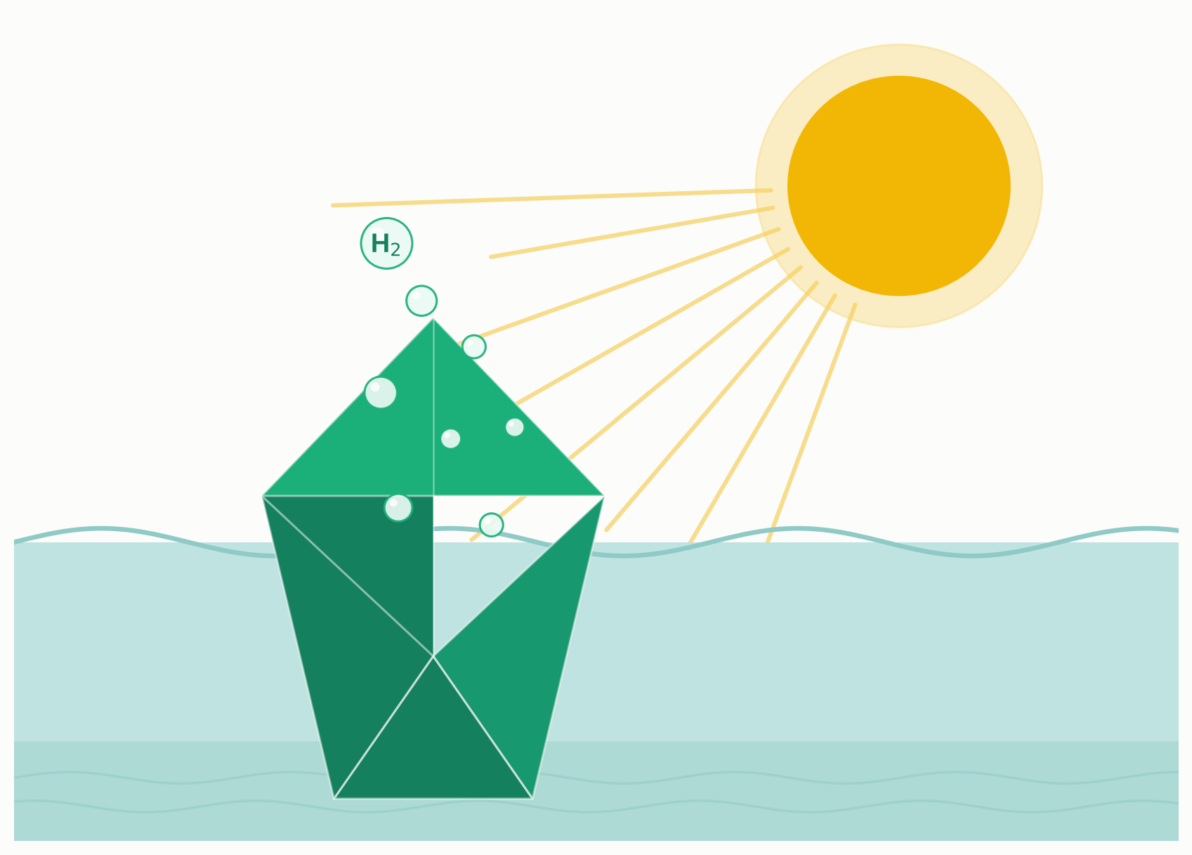
    Illustration of sunlight striking a crystal submerged in water, releasing hydrogen bubbles

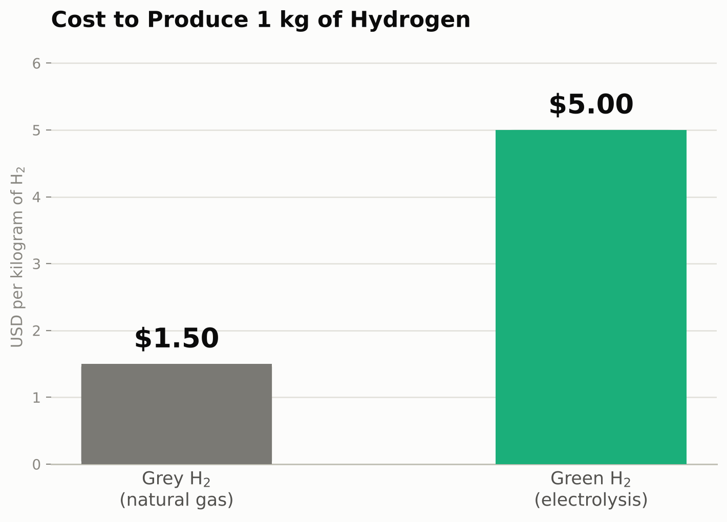
    Hydrogen is widely discussed as a fuel for ships, heavy trucks, steelmaking, and chemical plants that are hard to run on batteries alone, since it can generate electricity in a fuel cell with water as the main byproduct. The catch is how the hydrogen gets made. Today, hydrogen production is dominated by fossil fuels, mostly natural gas processed through a method called steam-methane reforming, with coal gasification also contributing a significant share, particularly in parts of Asia. That hydrogen is relatively cheap, on the order of $1.50 per kilogram by Oregon State’s figures, but it releases carbon dioxide in the process, which undercuts much of the climate case for switching to hydrogen in the first place.

    Hydrogen made by splitting water with renewable electricity or sunlight, often called green hydrogen, avoids that emissions problem, but it typically costs around $5 per kilogram today by the same estimate, more than three times as much. These are illustrative, broad figures rather than fixed prices; actual costs swing with local energy prices, plant scale, and technology. Still, part of the gap comes down to hardware: some electrolyzers and photocatalytic systems rely on precious-metal catalysts, platinum among them, to drive the reaction fast enough to be practical, and those materials are not cheap.

    Catching Light With Sulfur, Not Metal

    The Oregon State team, led by chemist Kyriakos Stylianou of the university’s Materials Discovery Laboratory, built a metal-organic framework, a class of crystalline, extremely porous materials assembled from metal ions linked together by organic molecules, that they call BVR-19. It forms spontaneously in water at room temperature, a synthesis route the researchers say avoids the high heat or pressure many materials need, though how that translates into a final production cost has not been established.

    The structural feature that matters most sits inside BVR-19’s organic linkers: a bond between two sulfur atoms that briefly breaks apart when struck by light, generating short-lived, highly reactive sulfur species. According to Oregon State’s announcement, it is these organic, sulfur-containing building blocks that help capture light energy and move electrons to where hydrogen is ultimately produced, working alongside the framework’s metal centers rather than leaving the metal to do that job alone.

    Cost comparison of grey and green hydrogen production, per kilogram in USD
    Source: approximate figures cited alongside Oregon State University’s announcement

    Metal-organic frameworks have drawn growing interest across chemistry over the past two decades because they are so tunable: change the metal, change the organic linker, and the pore size, surface area, and chemical behavior of the resulting crystal can shift dramatically, all within the same basic scaffold. That tunability is also what let the Oregon State team probe BVR-19’s inner workings, discussed next.

    Skipping the Bolted-On Metal Catalyst

    Photocatalytic water splitting conventionally needs more than a light-absorbing material: it usually also requires a separate metal co-catalyst, often platinum, physically attached to the structure to help the hydrogen-forming step run efficiently. Oregon State’s announcement describes BVR-19 as needing no additional expensive metal catalyst for that job, since its organic sulfur chemistry already contributes much of the light capture and electron transfer that a bolted-on catalyst would otherwise be doing.

    That should not be read as “metal-free.” BVR-19 still contains a metal center as part of its crystalline framework, and the team found that changing which metal sits there measurably changed how well the material performed, meaning the metal is far from irrelevant to the chemistry. The real, narrower claim is that no extra, separately added metal catalyst has to be layered on top of the base material the way platinum often is in other systems. It is a distinction worth keeping straight, because the innovation here is removing an added manufacturing step and its cost, not removing metal chemistry from the picture entirely.

    From One Experiment to Design Rules

    To understand why some versions of the framework outperformed others, the team systematically swapped out the metal ion at BVR-19’s core while leaving the rest of the structure essentially unchanged, then compared how each version performed under light. That side-by-side comparison let them connect differences in hydrogen output to specific changes in the material’s electronic structure, giving them a mechanistic explanation for why certain versions work better rather than only an empirical ranking of which ones do.

    “Our work provides a blueprint for designing better materials that can bring down the cost of green hydrogen,” Stylianou said in the university’s announcement of the findings. The team frames the result as a design principle to test and build on in future materials, rather than a finished, optimized catalyst.

    Research groups elsewhere are taking a similar mechanism-first approach to other energy-relevant reactions. A Kyoto University-led team, for instance, recently worked out in detail why a molybdenum molecular catalyst can convert nitrogen from the air into ammonia under comparatively mild laboratory conditions. In both cases, the advance was not a brand-new chemical but a clearer picture of why an existing catalyst behaves the way it does, which is usually what lets chemists improve a material on purpose instead of by trial and error.

    What Cheaper Hydrogen Could Unlock

    None of this means cheap, catalyst-light green hydrogen is imminent. Oregon State’s announcement and the peer-reviewed paper describe a laboratory-scale material and a design principle, not a measured hydrogen production rate, an efficiency figure, or a path to manufacturing; those numbers were not disclosed publicly alongside the findings. The work has not been tested at anything resembling industrial scale, and getting from a promising crystal in a lab to a working photocatalytic reactor typically requires years of further development, stability testing, and engineering, assuming it clears each step at all.

    What the study adds is a mechanistic foothold: evidence that letting an organic, sulfur-based structure share the light-absorbing and electron-transfer work can reduce a material’s dependence on costly bolted-on metal catalysts, at least in this framework, plus a method for figuring out which structural tweaks help and which do not. Whether that translates into cheaper hydrogen depends on factors the current study does not address, including the material’s hydrogen production rate, its durability over repeated use, and the cost of the reactor system built around it. If those later steps go well, incremental advances like this one, chipping away at the cost of individual components rather than reinventing hydrogen production overnight, are generally how energy technologies get cheaper over time. That is also what would let industries that are hard to electrify, such as steelmaking, shipping, and heavy trucking, lean more on low-carbon hydrogen instead of the fossil-derived hydrogen most of them use today.

    The project was funded by the Murdock Charitable Trust, the National Science Foundation, and Oregon State’s College of Science. For now, the result lives in a peer-reviewed paper and a university announcement, with the follow-up work, refining BVR-19, measuring its efficiency, and testing its durability, still ahead.

    Sources: Oregon State University, Journal of the American Chemical Society (DOI), ScienceDaily, Interesting Engineering

  • [Science] MIND Diet Linked to Years of Slower Brain Aging, 12-Year Study Finds

    [Science] MIND Diet Linked to Years of Slower Brain Aging, 12-Year Study Finds

    A diet designed around brain health may actually be protecting it. A 12-year study of nearly 1,650 American adults found that people who more closely followed the MIND diet — a hybrid of the Mediterranean and DASH diets — showed significantly slower shrinkage in brain regions tied to memory and thinking. Researchers calculate the effect as equivalent to roughly 2.5 years of delayed brain aging. The findings, published in the Journal of Neurology, Neurosurgery & Psychiatry, offer some of the strongest long-term evidence yet that everyday food choices in midlife may shape how the brain ages decades later.

    Inside the MIND Diet Study

    The MIND diet (Mediterranean-DASH Intervention for Neurodegenerative Delay) scores people from 0 to 15 based on how often they eat foods linked to brain health, such as leafy greens, berries, nuts, whole grains, fish, and olive oil, while subtracting points for red meat, fried food, sweets, cheese, and butter. For the new study, researchers from Zhejiang University, Harvard T.H. Chan School of Public Health, and the long-running Framingham Heart Study followed 1,647 adults from the Framingham Offspring Cohort, who were around 60 years old on average when the study began.

    Starting in the early 1990s, participants filled out detailed food-frequency questionnaires to calculate their MIND diet scores, with an average score of just under 7 out of 15 — meaning most people in the study were eating a moderately, not perfectly, brain-healthy diet. From 1999 onward, each participant underwent brain MRI scans roughly every few years — three scans on average — allowing researchers to track real changes in brain volume over a median follow-up of 12.3 years, rather than relying on a single snapshot. This repeated-measurement design is part of what makes the study notable: most diet-and-brain research relies on a single MRI scan compared against a single diet survey, which can only show a correlation at one point in time. Tracking the same people’s brains over more than a decade let the researchers instead measure the pace of change itself.

    Years of “Younger” Brain Structure

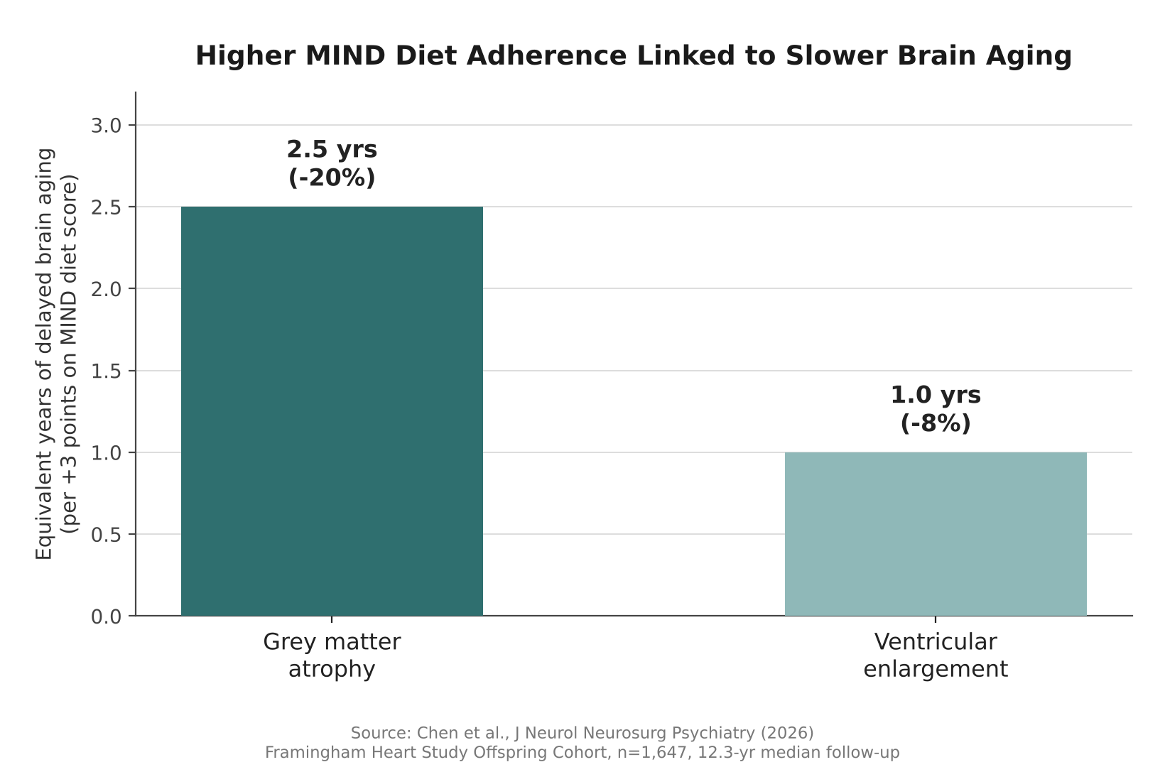
    The results were striking for an observational study. For every 3-point increase in MIND diet score, participants’ grey matter — the brain tissue that handles memory, decision-making, and sensory processing — shrank 0.279 cubic centimeters less per year, about 20% less age-related loss than would otherwise be expected. Researchers translated that gap into real-world terms: roughly 2.5 fewer years of brain aging.

    A 3-point higher MIND diet score was linked to about 2.5 fewer years of brain aging in grey matter volume — one of the clearest signals yet connecting everyday diet to long-term brain structure.

    Higher MIND scores were also linked to slower enlargement of the brain’s ventricles, fluid-filled spaces that expand as surrounding tissue is lost with age. That slower expansion — about 8% less than expected — translated to roughly one additional year of more youthful brain structure. Because all participants’ brains shrank somewhat over the study period, as is normal with aging, the real finding is about the pace of that decline, not whether it happened at all.

    Bar chart showing MIND diet score linked to years of delayed brain aging
    Source: Based on data from Chen et al., J Neurol Neurosurg Psychiatry (2026)

    Foods That Helped, and Surprised

    Not every food in the MIND diet scoring system behaved as expected once researchers looked food by food. Berries were linked to slower enlargement of the ventricles, and poultry was linked to both slower ventricular enlargement and slower grey matter loss — consistent with berries’ high antioxidant content and poultry’s role as a lean protein source, both of which may reduce oxidative stress that damages neurons over time.

    On the other end, sweets and fried fast food were linked to faster ventricular expansion and greater shrinkage of the hippocampus, the brain region most closely tied to memory formation. The researchers pointed to inflammation, unhealthy fats, and advanced glycation end-products — compounds that form when sugar reacts with proteins or fats in food, and which have been separately linked to tissue aging throughout the body — as possible mechanisms.

    Two results, however, ran against expectations. Whole grains, which earn positive points in the standard MIND scoring system, were associated with several unfavorable brain changes in this particular cohort, while cheese, which typically loses points, was linked to favorable ones. The study’s authors flagged both findings as unexpected and said they would need further research to explain, rather than drawing firm conclusions from them. One possibility researchers did not rule out is that people’s reasons for eating more or less of a given food — a doctor’s recommendation to cut dairy after a cardiovascular diagnosis, for instance — could confound a simple food-by-food analysis in ways a single study cannot fully untangle.

    Who Benefited Most

    The association between MIND diet adherence and slower brain aging was not uniform across the group. It was strongest among older participants, who face a faster baseline pace of brain aging and may have more to gain from protective factors. It was also stronger among people who were physically active and those who were not living with obesity, hinting that diet may work alongside — rather than instead of — other healthy habits.

    Participants who ate closer to the MIND pattern also tended to share other characteristics: they were more likely to be women and college-educated, and less likely to smoke or to have type 2 diabetes, high blood pressure, or cardiovascular disease. The researchers statistically accounted for these differences in their analysis, but they illustrate how diet quality tends to travel alongside a broader set of health behaviors.

    What This Study Can’t Prove

    Because this is an observational study, it cannot prove that the MIND diet alone causes slower brain aging. People who eat more healthily in general also tend to differ from others in exercise habits, education, and underlying health conditions, and some of the measured benefit may reflect those overlapping factors rather than diet in isolation. The food data also relied on questionnaires completed in the 1990s, which depend on participants’ memory and are vulnerable to reporting error, and the study could not fully rule out mild cognitive impairment at participants’ first brain scan or track how their diets may have changed over the following two decades.

    The study population was also overwhelmingly White, drawn from a single long-running cohort in Massachusetts, so it remains unclear how well the findings would generalize to more diverse populations. Still, the researchers conclude that their results “reinforce the potential of the MIND diet as a brain-healthy dietary pattern.” Diet is only one piece of the brain-aging puzzle — other recent work, including a study on how the Alzheimer’s risk gene APOE4 scars brain blood vessels, points to genetic and vascular factors working alongside everyday habits. Taken together, the evidence suggests that what people eat in midlife is worth taking seriously as a long-term investment in cognitive health, even before a formal clinical trial settles the question of cause and effect.

    Sources: BMJ Group, Journal of Neurology, Neurosurgery & Psychiatry (PMC)

  • AI Vision Misses an Illusion the Brain Shows — A Gap Worth Testing

    AI Vision Misses an Illusion the Brain Shows — A Gap Worth Testing

    A new study finds that after watching something move, people briefly misjudge exactly where a now-still object is sitting — and a matching shift shows up in signals recorded from monkeys’ brains. The seven AI vision models the researchers tested, built to mimic human sight, did not reproduce that shift at all. Published October 2, 2026 in the journal Current Biology, the work from York University in Toronto does not prove that this gap causes real-world AI vision systems to fail. What it does offer is a precise, repeatable benchmark that engineers building self-driving cars, robots, and other systems that must track a changing scene could use to check whether their software handles recent visual history the way biological vision does.

    Static-image benchmarks, how often a system correctly names or locates an object in a single photo, have long been a mainstay of computer-vision research. But the study’s authors point out that real cameras rarely see the world as a single frozen frame; they see a stream of images shaped by whatever happened a moment earlier. Notably, even a video-based network in the test, built specifically to use multiple frames over time, did not reproduce the shift seen in biological vision. That suggests the gap is not simply about feeding AI more frames, but about a specific kind of recalibration that today’s architectures do not learn on their own.

    A Trick of Motion Fools the Brain

    The effect the researchers studied is related to the classic motion aftereffect, often called the waterfall illusion because it was first described by people who stared at flowing water. After watching motion in one direction — stripes sliding left, for example — a stationary object shown afterward can appear displaced in the opposite direction, even though its pixels have not moved at all. The York University team used this positional bias as a precise, measurable probe into how the brain represents where an object is, rather than treating it as a mere curiosity.

    Testing Monkeys, Humans, and Machines

    The research team, led by senior author Kohitij Kar and first author Elizaveta Yakubovskaya, ran related versions of the same basic test on three very different visual systems. Seventy-nine human volunteers, 35 in a no-motion baseline group and 22 in each of two adaptation-direction groups, briefly saw an object flash on screen for a tenth of a second and clicked where they thought it was. Two rhesus macaques viewed similar sequences while chronically implanted electrode arrays recorded signals from more than 150 neurons in the inferior temporal (IT) cortex, a region of the brain’s object-recognition pathway whose activity also happens to carry information about where an object sits. The same images were then fed into seven widely used computer-vision models, ranging from classic image classifiers such as AlexNet and VGG-16 to a vision transformer and a video-based action-recognition network called SlowFast.

    AI Could Find Objects, Not the Illusion

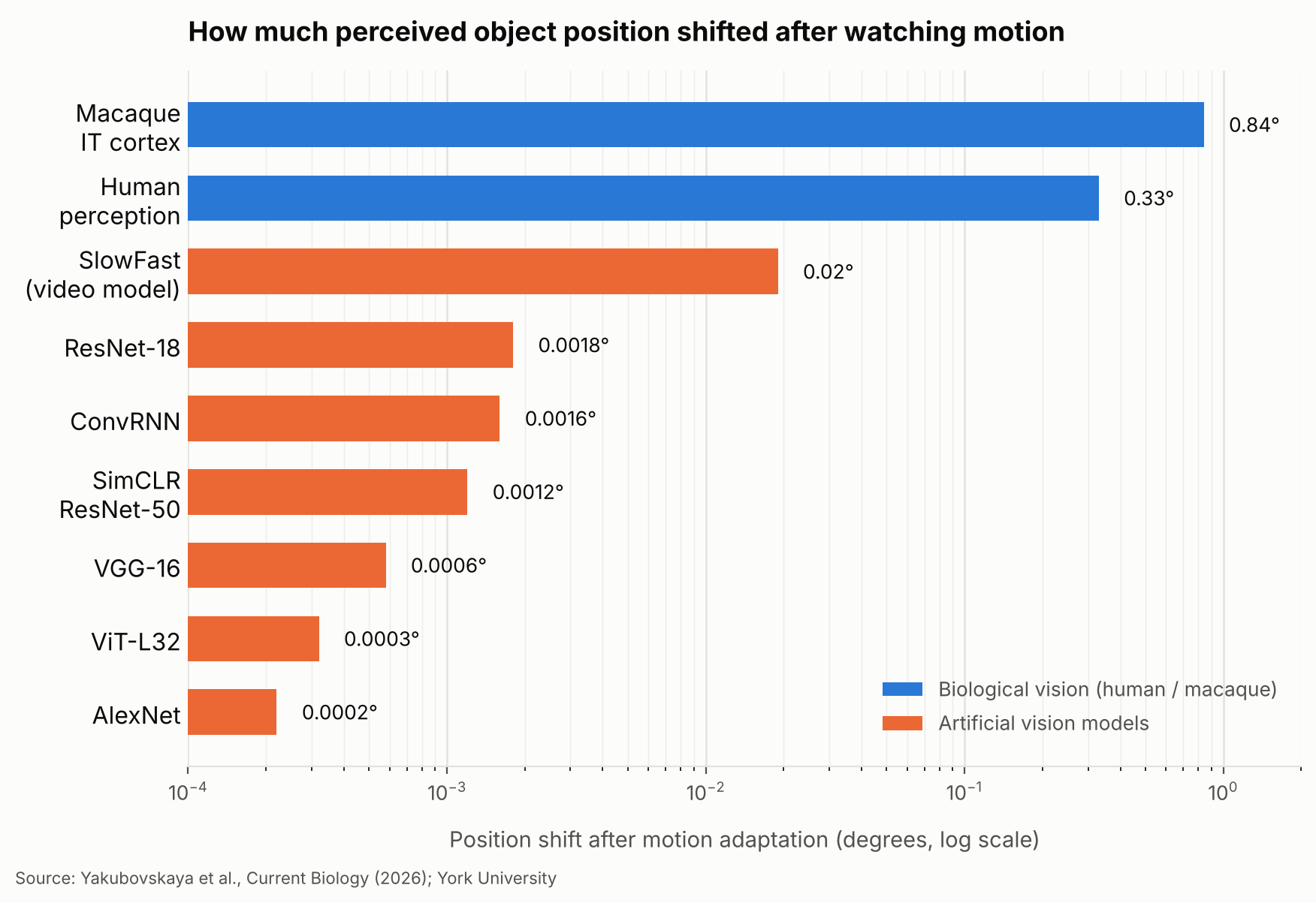
    On ordinary, non-adapted images, both the brain recordings and the AI models could track an object’s true horizontal position: linear readouts from several of the networks correlated with true position about as strongly as readouts from the monkeys’ own IT neurons did, in the range of roughly 0.59 to 0.84. So the models were not simply bad at locating objects. The difference showed up only after adaptation. Human volunteers reported an object’s position shifted by an average of 0.33 degrees of visual angle, opposite the direction of the preceding motion. The position decoded from the monkeys’ IT population activity shifted even further, by 0.84 degrees in the same direction, though that neural shift is a decoded signal rather than a direct report of what the animal consciously saw. Every AI model tested, including SlowFast, shifted by two-hundredths of a degree or less, in practice no meaningful shift at all.

    Human observers reported their perception shifting by 0.33 degrees after watching motion, and the position decoded from monkeys’ IT neurons shifted by 0.84 degrees — while every AI vision model tested shifted by two-hundredths of a degree or less.
    Bar chart comparing object-position shift after motion adaptation across human perception, macaque IT cortex, and seven AI vision models, on a log scale in degrees.
    Source: created based on data from Yakubovskaya et al., Current Biology (2026).

    Copying the Brain’s Adaptation Trick

    The gap did not mean the AI models were incapable of representing position-shifting information at all. When the researchers applied a mathematical transformation, derived from how IT neurons’ responses changed during adaptation, directly onto the models’ internal feature representations, the models began producing human-like position shifts. That shows the models’ feature spaces can support the effect once an appropriate transformation is imposed from outside. It does not show that any of the networks learned to generate that adaptation on their own during ordinary training, which is the gap the authors highlight. This is not the first time researchers have argued that borrowing the brain’s own computations, not just its final answers, could inform AI design; a separate study on how the brain re-files decision options made a related case for mining neural codes rather than just outputs.

    A Gap Worth Testing, Not Yet a Flaw

    The researchers are careful to frame this as a demonstrated difference in representations, not proof that AI vision performs worse in practice. Still, they argue it is a difference worth testing. Self-driving cars, delivery robots, and surveillance systems all judge where things are from a stream of frames in which recent motion could, in principle, matter. Consider a robot tracking a cyclist crossing its camera’s field of view: researchers could test whether the robot’s position estimate shifts the way a human’s perception would after similar recent motion, even though the current image alone has not changed. The York study does not show that reproducing this illusion would make such a system safer or more accurate; it offers a concrete, repeatable way to ask the question. Kar has argued more broadly that judging AI only by whether it reaches the right final answer can miss gaps like this one, and that matching the computations behind human perception, not only matching outputs, may help researchers evaluate whether an AI vision system handles a changing scene the way people expect.

    What the Study Doesn’t Yet Show

    The authors are explicit about the limits of their work. The experiments involved only two macaques and one specific illusion, so it is unclear how far the pattern generalizes to other forms of perceptual adaptation, to more animals, or to other stages of the visual pathway beyond IT cortex. The AI models tested were standard research networks for image and video recognition, not the newer multimodal AI assistants that combine vision with language, which the study did not evaluate. The researchers also do not claim that the perceptual bias they measured is actually beneficial for tracking objects in everyday life, only that it reflects a real computation in primate vision that current AI architectures do not reproduce on their own. They suggest the motion aftereffect could now serve as a simple, repeatable benchmark that other labs and companies use to test how their own vision models handle a changing visual world, and Kar’s group is already pursuing that direction, building what it calls more primate-like artificial neural networks for tasks such as continuous object tracking.

    Sources: Yakubovskaya et al., “The macaque IT cortex but not current artificial vision networks encode object position in perceptually aligned coordinates,” Current Biology (2026), Yakubovskaya et al., arXiv preprint (2026), York University press release coverage, via Neuroscience News

  • A Cancer Drug Already Tested in Patients Was Mainly Hitting a Different Target Than Scientists Thought — And That Could Open a New Path for Brain Cancer Treatment

    A Cancer Drug Already Tested in Patients Was Mainly Hitting a Different Target Than Scientists Thought — And That Could Open a New Path for Brain Cancer Treatment

    • A drug already tested in cancer patients turns out to work mainly by blocking a different target than scientists believed — a mix-up that could change how its past and future trials are read.
    • Researchers at the University of Sydney were testing whether the drug, called zavondemstat, could also work against glioblastoma, a particularly hard-to-treat brain cancer, when its effects stopped matching its supposed target, a protein family called KDM4.
    • Using brain-tumor cells grown from patients, genetic tricks, and detailed chemistry, the team traced the drug’s real effect to an enzyme called DHODH, which fast-growing cancer cells need to build new DNA.
    • The work is still confined to lab-grown cells and mice, not patients, but it also points to a possible new treatment route for glioblastoma, since other drugs that block the same enzyme already exist.

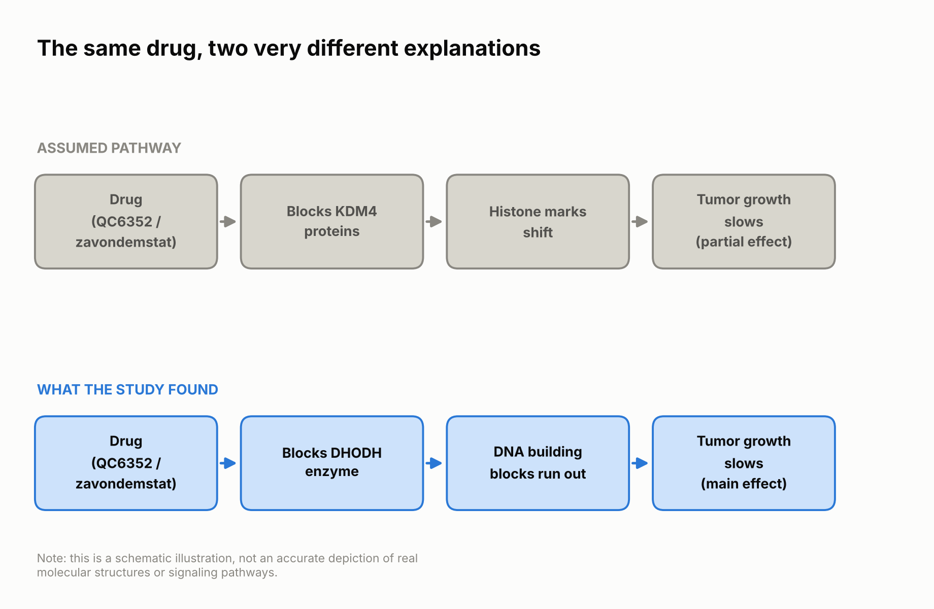
    Most new cancer drugs are designed around a specific molecular target: a protein that, when blocked, is expected to slow or stop tumor growth. But a study published on October 6, 2026 in Nature Chemical Biology by a University of Sydney-led team shows that an experimental drug already tested in cancer patients may owe most of its power to a completely different target than the one it was built for. The discovery does not mean the drug fails to work — it still slows cancer growth in the lab and in mice. What changed is the explanation for why, and that explanation matters for how the drug’s clinical trials — past and future — are designed and interpreted.

    A drug built for other cancers, tested against a tougher one

    The drug at the center of the study is called zavondemstat. It has already been tested in a phase 1 clinical trial of 30 patients with advanced, heavily pretreated solid tumors — about 40% of them colorectal cancer, along with prostate, pancreatic, and several other cancer types. Roughly 44% of the patients who could be evaluated had their disease stay stable on the drug, including a few who stayed stable for six months or more, and reported side effects were mild. The trial’s sponsor chose to halt the study early for business reasons before testing higher doses, so what comes next for zavondemstat is not yet settled. The drug was developed to block a family of proteins called KDM4, which many tumors produce in excess to help themselves grow and spread. A closely related laboratory compound, QC6352, has been widely used by researchers as a tool for studying what KDM4 does inside cells.

    A team led by Professor Lenka Munoz at the University of Sydney’s Charles Perkins Centre, working with collaborators at Goethe University, the University of Oxford, and the Institute of Cancer Research in London, wanted to see whether zavondemstat or QC6352 could also help against glioblastoma. Glioblastoma is the most common malignant brain tumor in adults and one of the hardest cancers to treat, so researchers routinely test whether drugs built for other cancers might also work against it.

    The target did not add up

    QC6352 showed strong activity against glioblastoma stem cells — tumor cell populations, grown from patient samples, that have stem-cell-like properties and are thought to drive the cancer’s growth and resistance to treatment. It blocked their growth at very low concentrations in lab dishes, while normal human brain support cells kept growing almost normally even at roughly ten times that concentration. That looked like confirmation that blocking KDM4 was the key to the effect. But when the researchers tested other drugs that block KDM4 just as strongly, those drugs failed to reproduce QC6352’s cancer-slowing effect. If KDM4 were really the main target, a different KDM4 blocker should have worked about as well. It did not, and that mismatch was the first sign that something else was going on.

    Tracing the main target: an enzyme called DHODH

    To find the true cause, the team ran a series of genetic and chemical tests. One enzyme kept coming up: DHODH, short for dihydroorotate dehydrogenase. DHODH helps cells manufacture pyrimidines, chemical building blocks that cells assemble into the nucleotides needed to make new DNA and RNA. Fast-dividing cancer cells depend heavily on a steady supply of these building blocks, so blocking DHODH can slow their growth sharply.

    Two lab experiments made the case convincingly. First, the researchers added extra uridine, a related building block that cells can absorb directly and use to bypass a blocked DHODH; this restored the cells’ growth despite the drug still being present, which is a sign that DHODH, not KDM4, was what the drug was really acting through. Second, when they engineered cancer cells to carry a version of DHODH that the drug could not grab onto, those cells also kept growing normally even with the drug present. Both are standard laboratory rescue experiments, not something that could be done safely in a patient, but together they pointed squarely at DHODH as the main driver of the anti-cancer effect.

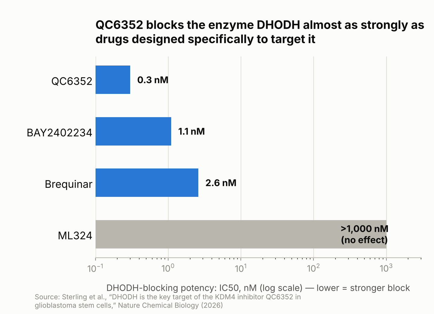
    The chemistry backs this up. In a test-tube enzyme assay, QC6352 blocked DHODH at a concentration of about 0.3 nanomolar — an extremely small amount — which is in the same range as drugs built specifically to target DHODH, such as brequinar (2.6 nanomolar) and BAY2402234 (1.1 nanomolar). A true KDM4-only inhibitor called ML324, by contrast, barely touched DHODH at all even at concentrations over a thousand times higher. These numbers describe how tightly each compound grabs the enzyme in a dish, not how well it would work in a patient, but the gap between QC6352 and ML324 is the clearest sign that QC6352’s anti-cancer effect runs mainly through DHODH. The researchers now describe zavondemstat and QC6352 as “dual KDM4–DHODH inhibitors”: both targets are blocked, but the DHODH side appears to be doing most of the work against glioblastoma.

    Schematic diagram comparing the assumed drug pathway through KDM4 with the pathway the study found through DHODH
    Scientists assumed the drug worked mainly by blocking KDM4 (top). The study found it mainly works by blocking DHODH instead (bottom). Schematic illustration, not an accurate depiction of real molecular structures.
    Bar chart comparing DHODH-blocking potency of QC6352, BAY2402234, brequinar, and ML324
    In a test-tube enzyme assay, QC6352 blocked DHODH about as strongly as drugs designed specifically for that purpose, while a KDM4-only drug barely blocked it at all. Data: Sterling et al., Nature Chemical Biology (2026).

    Why this matters beyond the lab

    QC6352 has been used as a standard research tool in laboratories around the world to study what KDM4 does. If much of its effect actually comes from DHODH, some of those earlier studies may need to be re-examined, since what looked like evidence about KDM4 biology could instead reflect DHODH biology.

    There is a more immediate concern, too. Zavondemstat has already been given to patients in a clinical trial that was designed and interpreted around the idea that it works mainly through KDM4. Professor Munoz has pointed out that getting a drug’s mechanism wrong is not just a historical curiosity — it can lead to poorly designed trials, choosing the wrong patients to enroll, and wasted research effort, and she argues this kind of mix-up is still common in cancer drug development today. To help avoid repeating the problem, her team also designed new experimental compounds that block KDM4 without touching DHODH at all, giving future researchers cleaner tools to study KDM4 on its own.

    A possible upside for brain cancer patients

    The mix-up also comes with a possible silver lining. In mice carrying human glioblastoma tumors, QC6352 extended median survival from 28.5 days to 34.5 days — an absolute difference of six days, or about 20% longer than in untreated mice — while normal brain support cells tolerated roughly ten times the dose without obvious harm in lab tests. That is an early signal of activity in animals, not evidence of a survival benefit in people. Still, because several drugs that block DHODH already exist and are being studied for other cancers, this discovery could give researchers a head start on exploring DHODH-targeted treatment for glioblastoma specifically, rather than starting drug development from zero. Much work would still be needed to show that an existing DHODH inhibitor can reach brain tumors at a safe dose and actually help patients.

    In mice, blocking the enzyme DHODH with QC6352 stretched median survival from 28.5 to 34.5 days — six days, or about 20% longer than no treatment at all.

    It is worth being careful about how far this points, though. The findings so far come only from lab-grown cells and mice, not from glioblastoma patients, and zavondemstat’s one completed human trial was not designed with DHODH in mind. Researchers elsewhere are also exploring other ways to make cancer treatment more targeted and less damaging to healthy tissue, such as an approach that gets cancer cells to manufacture their own chemotherapy drug while sparing normal cells. The DHODH finding adds another option to that broader search: not a cure in hand, but a clearer, evidence-based reason to try a treatment path that already has other drugs ready to test.

    Sources: University of Sydney, Nature Chemical Biology

  • How Much Are the World’s Oceans Really Warming? A New Method Finds a Large Gap With a Widely Used Climate Estimate — and Heat Moving Unevenly Between Ocean Basins

    How Much Are the World’s Oceans Really Warming? A New Method Finds a Large Gap With a Widely Used Climate Estimate — and Heat Moving Unevenly Between Ocean Basins

    • Almost all of the extra heat trapped by human-caused warming ends up in the ocean, so how well we can measure that heat shapes how much we can trust climate tracking and forecasts of rising seas.
    • German researchers built a new way to estimate that heat, combining data from floating ocean sensors and two kinds of satellites to recalibrate the computer reconstructions scientists normally rely on.
    • For part of the past two decades, one widely used reconstruction estimated far more ocean heat uptake than the new study did — in one stretch, nearly double.
    • The heat also hasn’t spread evenly: the Indian Ocean absorbed an outsized share between 2005 and 2015, a pattern that has since become more evenly spread across the Pacific, Indian, and Atlantic Oceans.

    Why the Ocean’s Heat Bill Matters

    Earth is currently taking in more heat from the sun than it radiates back out to space, mainly because greenhouse gases trap extra energy near the surface. Almost all of that surplus heat — roughly 90%, according to the new study — ends up in the ocean rather than in the air or on land. By absorbing so much of that excess energy, the ocean slows down how fast the atmosphere and land surface heat up, which is a large part of why it’s one of the most closely watched numbers in climate science.

    Scientists actually track two related but different numbers here: ocean heat content, meaning how much extra heat is already stored in the ocean, and ocean heat uptake, meaning how fast the ocean is gaining more of it, usually measured in watts per square meter of ocean surface. This story is mostly about the second number — the uptake rate, and how it differs from one ocean region to the next. As seawater heats up, it also expands slightly, a process called thermal expansion (think of a thermometer, where the liquid rises in the tube as it warms and takes up more space). That expansion drives a significant share of global sea-level rise.

    Getting a precise number for how fast the ocean is taking up heat, and where, turns out to be surprisingly hard. Researchers at the University of Bonn in Germany, in a study published in October 2026 in the journal Earth System Science Data, set out to make that accounting more reliable — and to see how it compares with the estimates climate scientists currently rely on.

    Three Data Sources, One Recalibrated Picture

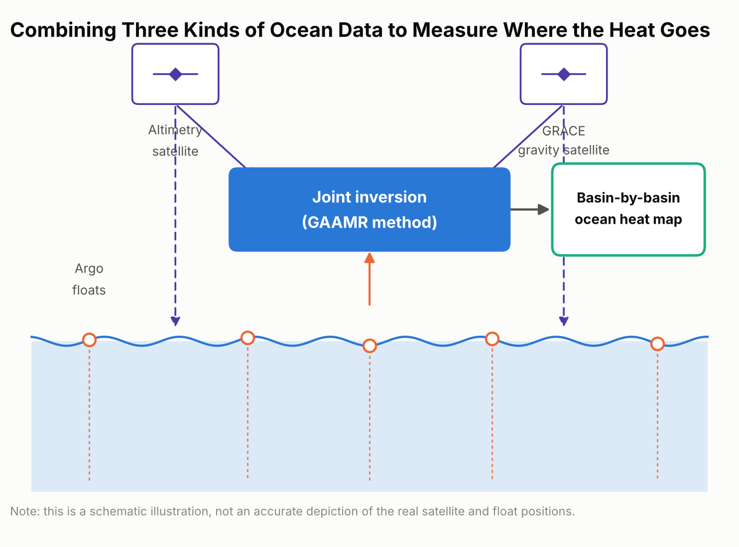
    No one can scoop up the ocean and measure its heat directly. Scientists instead combine indirect clues, and each one has blind spots on its own. The Bonn team’s new approach, which they call GAAMR (Grace, Altimetry and Argo Mode Rescaling), draws on three streams of satellite and ocean data: two satellite-based measurements and one array of ocean-going floats.

    The first is a global fleet of thousands of drifting robotic sensors called Argo floats, which sink and rise through the upper two kilometers of the ocean roughly every ten days, recording temperature and salinity as they go; Argo samples the upper ocean far more densely than the deep ocean, so deeper water is handled differently, as explained below. The second is satellite radar altimetry, which tracks the height of the sea surface from orbit; averaged over time, it can measure global sea-level trends to within a couple of millimeters a year, and warmer water takes up more space, so part of that rise reflects ocean expansion. The third is data from GRACE, a pair of satellites that detect tiny shifts in Earth’s gravity field caused by moving mass on and near the surface, including ice sheets, glaciers, and water stored on land. That gravity data helps researchers separate the part of sea-level rise caused by this extra mass from the part caused by the ocean simply expanding as it warms.

    Importantly, the new method isn’t a model-free measurement. It still starts from an ocean model — the same ORAS5 reanalysis used for comparison below — to map out the likely regional patterns of warming. What’s different is that, rather than trusting those modeled patterns at face value or applying one global adjustment factor to the whole ocean, the researchers rescale the patterns region by region using the real satellite and float observations. That regional rescaling, instead of a single worldwide correction, is the method’s main innovation, and it produces monthly, region-by-region estimates of ocean heat uptake rather than one rough global figure.

    Schematic diagram showing the three data sources combined in the GAAMR method: Argo floats, satellite altimetry, and GRACE gravity measurements
    The GAAMR method combines three data sources — Argo floats, satellite altimetry, and GRACE gravity data — to rescale ocean heat uptake estimates region by region.

    What the New Numbers Show

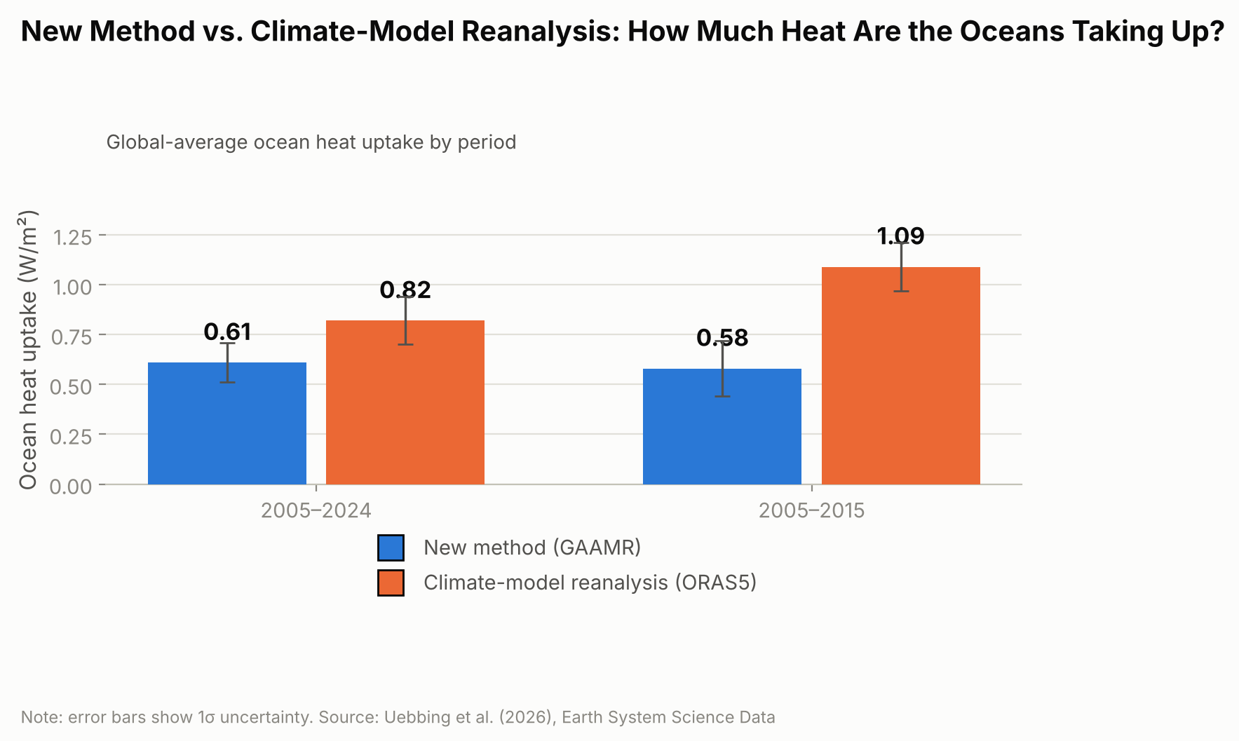
    Added up across the whole ocean, the new approach lines up reasonably well with several other independent estimates: it puts global ocean heat uptake from 2005 through 2024 at 0.62 watts per square meter, averaged over every patch of ocean surface on Earth. That figure is broadly consistent with estimates built from the Argo float network alone and with NASA’s CERES satellite measurements of Earth’s energy budget.

    Where the new estimate diverges is in comparison with ORAS5 specifically — a widely used “ocean reanalysis,” meaning a computer reconstruction that blends an ocean circulation model with whatever real-world observations are available, rather than a forward-looking climate projection. Over 2005–2024, ORAS5 put global ocean heat uptake at about 0.82 watts per square meter, roughly a third higher than the new study’s figure. The gap was much larger for the first half of that period.

    For 2005 to 2015, ORAS5 estimated ocean heat uptake at 1.09 watts per square meter, compared with 0.58 watts per square meter from the new GAAMR reconstruction — almost double.

    Bar chart comparing ocean heat uptake in watts per square meter from ORAS5 and the new GAAMR method for 2005 to 2024 and 2005 to 2015
    For 2005 to 2015 alone, the gap between ORAS5 and the new GAAMR estimate is nearly twofold.

    The researchers are careful not to frame this as proof that climate models in general overstate ocean warming. Their own result broadly agrees with several independent observational estimates; the clearer gap is specifically with ORAS5, suggesting that particular reanalysis may be overstating how fast the ocean has taken up heat, especially during the 2005–2015 decade.

    Heat on the Move: Why Some Ocean Basins Differ

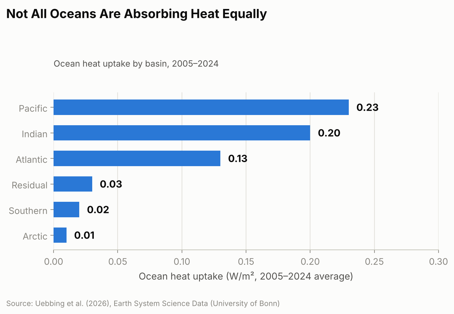
    The new regional estimates also reveal something a single global number can’t: heat hasn’t been spreading evenly across the world’s oceans. Over the full 2005–2024 period, the Pacific, Indian, and Atlantic Oceans together accounted for the large majority of the extra heat, with the Pacific absorbing the most, followed closely by the Indian Ocean and then the Atlantic. The Southern Ocean and Arctic Ocean, despite their size, contributed only a few percent combined.

    Horizontal bar chart showing the regional breakdown of ocean heat uptake by basin for 2005 to 2024, covering the Pacific, Indian, Atlantic, Southern, and Arctic Oceans and a residual term
    The Pacific, Indian, and Atlantic Oceans together account for the large majority of the added heat.

    That balance, though, looks more even over 20 years than it did at the start. From 2005 to 2015, the Indian Ocean absorbed heat at a disproportionately high rate compared with its share of global ocean area. One contributor researchers point to is ocean circulation: heat transport patterns linked to the Indonesian Throughflow, a current system that channels warm Pacific water through the gaps between Indonesia’s islands into the Indian Ocean, appear to have shifted after around 2018, with larger outflows of heat carrying it away from the equatorial Indian Ocean. The study doesn’t pin down the mechanism conclusively, but that shift coincides with the broader move toward a more even split among basins that is clear in the full 20-year record. Other consequences of the same underlying warming are easier to see because they happen on land you can visit — this year’s near-record glacier melt across the Swiss Alps (the second-largest annual loss on record) is one example — even though the ocean, out of sight below the surface, is where almost all of the extra heat actually ends up.

    Why Getting This Number Right Matters

    Thermal expansion from ocean warming accounted for an estimated 42% of global sea-level rise between 1993 and 2015, according to the Intergovernmental Panel on Climate Change, and considerably more in some regions, so an inflated or understated ocean-heat figure can throw off coastal planning that depends on sea-level projections. The Bonn researchers also argue that ocean heat content is a steadier gauge of global warming than surface air temperature, which can swing sharply from a single hot or cool year in a way the ocean’s vast, slow-moving mass does not.

    The approach is not a forecasting tool — it’s a more careful reconstruction of what has already happened, built from twenty years of historical satellite and float records. The team says extending it further back in time is limited mainly by how sparse the satellite and ocean-observing records become before the early 1990s, and they plan to incorporate upcoming missions, including proposed successors to GRACE such as GRACE-C and NGGM, to keep the record going. They also call for sustained, internationally coordinated funding for the ocean- and gravity-monitoring satellites the method depends on, warning that gaps in that coverage would make it harder to catch the next shift in where the planet’s extra heat is headed.

    Sources: University of Bonn, “How Much Are the World’s Oceans Warming?”; Uebbing et al., “Improving global and regional ocean heat content by consistently combining GRACE gravity, satellite altimetry, and Argo profile observations in a joint inversion framework,” Earth System Science Data, 2026.

  • [Science] China’s Chang’e-6 Moon Samples Reveal a Rare Magnetic Mineral — A New Lead in the Mystery of the Moon’s Lost Protective Field

    [Science] China’s Chang’e-6 Moon Samples Reveal a Rare Magnetic Mineral — A New Lead in the Mystery of the Moon’s Lost Protective Field

    • Earth’s magnetic field acts like an invisible shield, deflecting much of the charged-particle wind that streams from the Sun — but the Moon and Mars both lost a global magnetic field like this billions of years ago, and scientists still do not fully agree on why.
    • Researchers studying soil that China’s Chang’e-6 mission brought back from the Moon’s far side found a rare, naturally magnetic form of iron that had never before been confirmed in real Moon rock.
    • Using a specialized microscope, they showed that this iron can hold a stable magnetic swirl pattern — the kind of structure that can, in principle, lock in a trace of the magnetic field present when it formed.
    • This is an early-stage finding: it does not by itself settle the long-running debate about the Moon’s lost magnetic field, but it gives scientists a new kind of physical evidence to test their ideas against.

    A missing magnetic shield, and a Moon that might hold a clue

    Right now, a layer of invisible magnetism generated deep inside Earth is quietly doing one of the most important jobs for life on this planet: deflecting much of the charged-particle wind that streams from the Sun, known as the solar wind, so it does not gradually strip away our atmosphere. Mars does not have this shield anymore. Neither does the Moon. Both are thought to have had one billions of years ago, generated by a “dynamo” — a churning flow of molten or partly molten metal inside the planet that behaves like a natural electric generator. Figuring out when and why each dynamo switched off is one of the open questions in planetary science, partly because it is also a preview of how a rocky world’s interior can change over very long timescales.

    The Moon is a convenient place to study this, because unlike Mars, it is close enough that spacecraft have brought real samples home. In 2024, China’s Chang’e-6 mission did something no previous mission had: it collected soil from the Moon’s far side, from the Apollo basin inside the giant South Pole–Aitken impact basin. A team at the Hefei Institutes of Physical Science (HFIPS), part of the Chinese Academy of Sciences, has now reported a new discovery hiding inside that soil, published online September 16, 2026, in the Proceedings of the National Academy of Sciences (PNAS).

    An iron that “shouldn’t” exist — until an asteroid hit made it

    The team, led by Prof. Haifeng Du, was looking at tiny beads of impact glass: droplets of rock that melted and then froze solid within moments after an asteroid or other object slammed into the Moon’s surface. Scattered through that glass, they found nanometer-scale particles — far smaller than a speck of dust, ranging from under 10 nanometers to about 140 nanometers across — made of a form of iron called gamma-iron, or γ-Fe for short.

    Gamma-iron is not exotic chemistry; it is simply metallic iron arranged in a different crystal structure than the form most people picture. At ordinary pressure, pure iron normally settles into a structure called alpha-iron as it cools, and that is the arrangement found in most familiar iron-rich rocks. Gamma-iron is normally only stable over a high-temperature range, roughly 912°C to 1,394°C, and should flip back into the ordinary form well before reaching room temperature. Finding it “frozen” in place at room temperature, inside real Moon soil, was unexpected. The researchers propose that the extremely rapid cooling that follows a high-speed impact — far faster than rock cools under ordinary conditions — combined with the small amount of trace elements in the particles, trapped the iron in this unusual structure before it had a chance to transform.

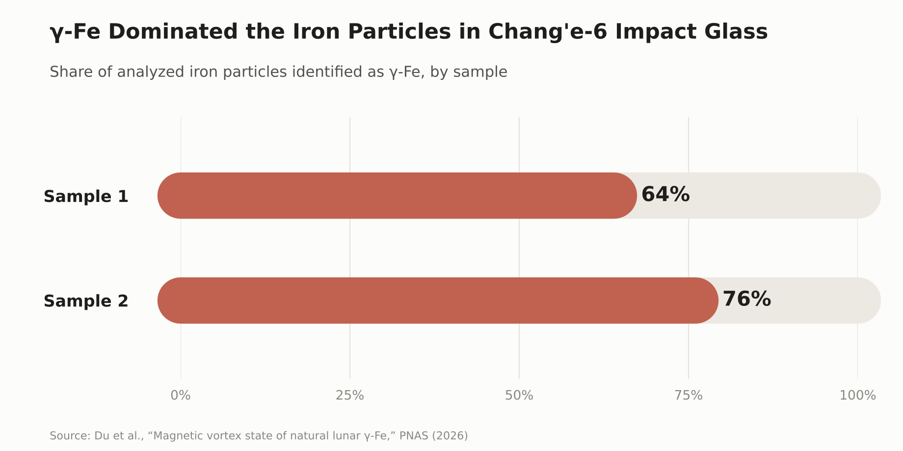
    Among 50 iron particles the team examined across two impact-glass samples, γ-Fe was the dominant crystal phase: it accounted for roughly 64% of the particles analyzed in one sample and 76% in the other. The particles also contained very little nickel, below 0.8% by weight, which helps distinguish them from the iron-nickel alloys already known from meteorites and other Moon rocks.

    Bar chart showing the share of analyzed iron particles identified as gamma-iron in two Chang’e-6 impact-glass samples: 64% and 76%.
    γ-Fe was the dominant iron phase in both impact-glass samples the team analyzed in detail.

    Reading a magnetic memory under the microscope

    Finding an unusual mineral is interesting on its own, but the more important part of the discovery is what the iron does magnetically. Using a specialized imaging technique called off-axis electron holography — a way of directly visualizing magnetic structure at a scale of billionths of a meter, inside an electron microscope — the team found that γ-Fe particles larger than about 100 nanometers settle into a stable “single-vortex” magnetic state: the magnetization inside the particle curls around in a stable loop, rather than pointing in one simple direction.

    That stability matters. A magnetic structure that holds its shape reliably, and responds in a predictable way to outside magnetic fields, is the kind of structure that can carry what scientists call remanent magnetization — a lasting trace of the magnetic field present when the material formed and cooled. In principle, that makes it somewhat like certain magnetic minerals in Earth’s own rocks, which preserve a record of our planet’s past magnetic field. Dr. Long Li, one of the researchers on the team, put it this way:

    “This tiny magnetic fossil may help us better understand the Moon’s ancient magnetic history.” — Dr. Long Li, Hefei Institutes of Physical Science

    It is worth being precise about what has, and has not, been shown so far. The team has demonstrated that γ-Fe exists in real lunar material, that it forms a stable magnetic vortex state, and that this state is capable of recording magnetization. What is not yet established is which magnetic field any particular particle actually recorded — the Moon’s ancient, planet-wide dynamo field, or a brief, local field generated by the heat and shock of the very impact that formed the glass bead around it. Telling those two signals apart is exactly the next step the researchers say this discovery opens up.

    A genuinely unsettled debate about the Moon’s lost magnetism

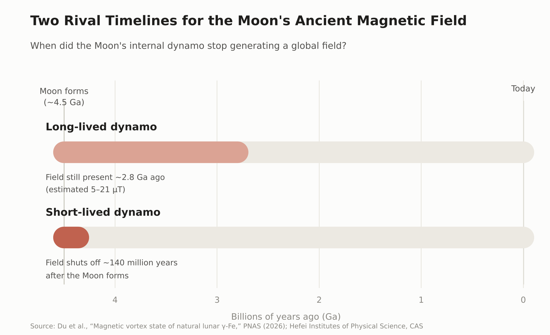
    Scientists already have evidence the Moon once had a working dynamo, because older Moon rocks brought back decades ago by the Apollo missions carry their own trace of ancient magnetism. Exactly how strong that field was, and especially how long it lasted, is still disputed — and this is a real, ongoing scientific disagreement rather than a settled fact with one outlier view. Paleomagnetic measurements of Chang’e-6 basalt reported a field of roughly 5 to 21 microtesla as recently as about 2.8 billion years ago, a result some researchers read as pointing toward a longer-lived dynamo. Other researchers, using different measurement methods on Apollo-era samples, have argued that the evidence for a long-lived global field is weaker than it first appeared, and that the core dynamo may have been largely confined to the Moon’s first 140 million years or so, with anything recorded afterward reflecting brief, impact-generated fields rather than a steady, planet-wide one.

    Timeline diagram comparing two rival hypotheses for when the Moon’s internal dynamo stopped generating a global magnetic field.
    Two competing timelines for when the Moon’s ancient dynamo shut off — a debate γ-Fe could eventually help settle.

    This is where γ-Fe could eventually contribute. Because it forms under different conditions than the more common alpha-iron, and appears specifically in impact-formed glass, comparing what it and other magnetic minerals “remember,” across many more samples, may eventually help researchers test whether a given signal reflects the Moon’s global dynamo, an impact-generated field, or some mixture of the two.

    Still early days: what comes next

    The team has not announced a new, confirmed timeline for when the Moon’s dynamo turned off, and γ-Fe on its own cannot provide a precise date for that event. What they have shown is that this magnetic mineral exists in real lunar material, that it can hold a stable magnetic state at the nanometer scale, and that it is a promising new tool for a field that has had to work with a frustratingly small and ambiguous set of physical clues. More broadly, understanding how a rocky world like the Moon or Mars generates, and eventually loses, its own internal magnetic field is part of a wider effort to understand what shapes a planet’s long-term climate and atmosphere — knowledge that also feeds into how scientists assess the conditions on rocky planets orbiting other stars. A small, newly identified mineral will not settle that larger question by itself, but it gives researchers one more concrete thread to pull on.

    Sources: Du, H. et al., “Magnetic vortex state of natural lunar γ-Fe,” PNAS (2026) · Hefei Institutes of Physical Science, CAS · ScienceDaily · The Brighter Side of News

  • [Science] Stimulating the Vagus Nerve After Practice Helps Mice Learn Better Days Later — Tohoku University Finds a Link to Rhythmic Changes in Brain Blood Volume

    [Science] Stimulating the Vagus Nerve After Practice Helps Mice Learn Better Days Later — Tohoku University Finds a Link to Rhythmic Changes in Brain Blood Volume

    • Applying electrical stimulation to the “vagus nerve” on the left side of the neck right after practicing something new may make mice better at it days later, a new study suggests.
    • If a similar effect holds in humans, it could eventually point toward new ways to support motor learning or rehabilitation.
    • Tohoku University researchers stimulated the vagus nerve while tracking local blood-volume changes in the brain, and found that repeated stimulation produced rhythmic fluctuations in local blood volume.
    • Mice with larger blood-volume oscillations after stimulation tended to score higher on a learning test five days later. This is a mouse study, though, and it’s not yet known whether the same effect would occur in humans.

    The Vagus Nerve — a Key Line Between Brain and Body

    The vagus nerve is a major nerve that runs from the brainstem through the neck, chest, and abdomen, carrying signals between the brain and organs such as the heart, lungs, and digestive tract. It’s one of the main nerves connecting the brain and internal organs in both directions: it carries signals about the body’s internal state — such as heart rate, breathing, and digestion — up to the brain, while also carrying signals from the brain back down to those organs.

    A treatment that stimulates this nerve from the outside with electrical pulses is called “vagus nerve stimulation,” or VNS. Using an implanted device, it’s an established therapy for epilepsy that doesn’t respond well enough to medication. In recent years, researchers have also been studying a non-invasive approach that stimulates part of the ear through the skin — though that’s a different method from the one used in this mouse study. Exactly what VNS changes inside the brain has remained unclear.

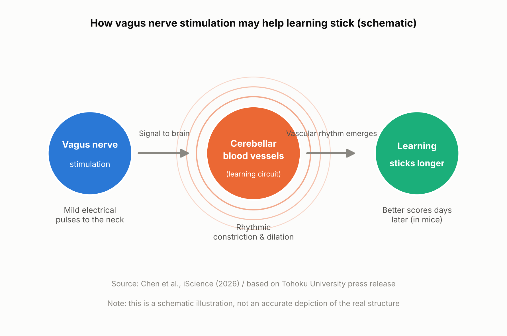
    Schematic showing how vagus nerve stimulation and a brain blood-volume rhythm relate to long-term learning
    How vagus nerve stimulation and a brain blood-volume rhythm relate to long-term learning (schematic illustration, not an accurate depiction of the real structure).

    The Mouse Experiment: Stimulation Came After Practice

    A team at Tohoku University’s Graduate School of Life Sciences — Junyu U. Chen, Assistant Professor Yoko Ikoma, and Professor Ko Matsui — fitted a small cuff electrode around the left cervical vagus nerve in mice and delivered electrical stimulation. The mice practiced a task called the horizontal optokinetic response (HOKR): tracking a vertical striped pattern that moved back and forth in front of them with their eyes. This kind of task is known to depend heavily on the cerebellum, a brain region central to motor learning.

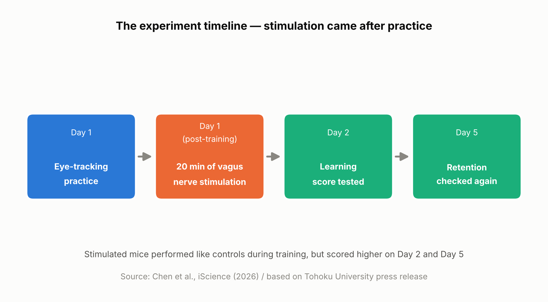
    On day one, the mice practiced for 15 minutes, four times, spaced an hour apart. Right after each day-one training session, mice in the stimulated group received 20 minutes of vagus nerve stimulation at one of two intensities, while control mice received none. Performance was then measured again on day two and day five.

    On day one itself, there was no difference between the stimulated and unstimulated mice. But by day two and especially day five, the stimulated mice showed higher scores, with the higher-intensity stimulation producing a visible benefit as early as day two and both intensities showing an advantage by day five. In other words, VNS didn’t directly boost performance during training — it appears to have improved later-day learning through some process that unfolded after training ended.

    Timeline of the mouse experiment, showing stimulation delivered after practice
    The experiment timeline. Based on Chen et al., iScience (2026) and the Tohoku University press release.

    Blood Volume Around the Cerebellum Fell Into Rhythm After Stimulation

    Using a light-based technique called fiber photometry, the researchers measured local blood-volume changes in tissue around the cerebellar flocculus. Each train of vagus nerve stimulation produced a two-step change: local blood volume briefly dipped, then — after a short delay — rose. This pattern is consistent with alternating contraction and dilation of blood vessels in that area.

    With repeated stimulation, this rise-and-fall pattern settled into a steady rhythm. The researchers think this rhythmic change in blood volume may be related to how the brain regulates local blood flow and its metabolic environment — but the study did not directly measure oxygen, ATP, or other metabolic markers, so exactly how the blood-volume rhythm might support learning is still unclear.

    Long-term learning is thought to involve various changes in the brain, including “synaptic plasticity” — changes in the connections and activity between neurons — which requires energy. The research team’s idea is that if the blood-volume rhythm is tied to how well-nourished brain tissue is, it could be helping support those changes, though this remains a hypothesis rather than something the study directly confirmed.

    Mice with larger blood-volume oscillations after stimulation tended to score higher on the learning test five days later

    A Bigger Rhythm, Better Long-Term Learning — but Only a Correlation So Far

    The most notable finding was a relationship between the magnitude of the blood-volume oscillations in the 20 minutes right after stimulation and performance five days later: mice with larger oscillations tended to score higher on day five. It’s worth being clear that this is a correlation, not proof that the blood-volume change caused the better learning.

    “The key point is that VNS was delivered only after training,” said Professor Matsui. “Our findings suggest that VNS may open a hidden window of opportunity for enhanced learning by making the brain environment more receptive to long-lasting change.” Lead author Junyu Chen added, “Our brains may be more strongly influenced by the body than we imagine. By tuning the brain’s metabolic environment, including rhythmic vascular movements, we may eventually unlock capacities that would otherwise remain latent.”

    What’s Still Unknown — and What Comes Next

    Based on this study alone, it’s not possible to say whether the blood-volume rhythm actually causes the learning improvement. Confirming that would require future experiments that directly manipulate blood-vessel or blood-volume activity and track the effect on learning. The researchers also did not directly measure metabolic molecules such as ATP, so the proposed mechanism remains a hypothesis.

    It’s also worth remembering that this was a mouse study using an invasive cuff electrode surgically placed around the cervical vagus nerve, delivering stimulation directly to the nerve rather than through the skin. Less invasive, ear-based stimulation methods are already being studied in humans for other purposes, but whether this mouse study’s results would carry over to humans — or to those non-invasive methods — can’t be determined from this result alone. The research team suggests that fine-tuning the timing and intensity of stimulation could eventually help support learning and rehabilitation, though that application remains a research goal rather than something available today.

    Sources: Tohoku University Graduate School of Life Sciences press release (August 26, 2026), Chen et al., iScience (published online August 25, 2026)

  • [Science] Scientists Build a Billion-Dalton Molecular Thread Longer Than a Cell — It Quadrupled Nerve-Cell Activity in the Lab

    [Science] Scientists Build a Billion-Dalton Molecular Thread Longer Than a Cell — It Quadrupled Nerve-Cell Activity in the Lab

    • A newly built, self-assembling material made nerve cells growing in a lab dish roughly four times more biologically active than usual, hinting that materials like it could one day help repair damaged nerves.
    • Chemists at Northwestern University found a new way to precisely control how long a certain kind of molecular thread grows, and how its positively and negatively charged segments are arranged along the way.
    • By molecular standards the resulting threads are enormous — roughly 100 times heavier than typical plastic-like polymers and bigger than even the largest proteins found in nature — even though they are built from many smaller molecules that click together on their own, rather than one molecule held together by ordinary chemical bonds.
    • So far the effect has only been shown in cells grown in a lab dish, not in animals or people, so any real treatment is still a long way off.

    Billion-Dalton Supramolecular Polymers: A Structure Big Enough to Rival a Cell

    A team led by the chemist Samuel I. Stupp at Northwestern University has built a new kind of giant structure, described in the journal Science on October 1, 2026. It belongs to a class of materials called supramolecular polymers. Rather than being a single molecule held together by strong chemical bonds the way an ordinary plastic is, it is made of many smaller molecules that click onto one another and hold themselves together more loosely, the way a line of magnets snaps together end to end. That looseness usually makes supramolecular polymers easy to assemble but hard to control, since there has been no reliable way to stop them from growing at a chosen point.

    Scientists weigh molecules in daltons, a unit named after the 19th-century chemist John Dalton, where one dalton is roughly the mass of a single hydrogen atom. Northwestern researchers point to common high-mass synthetic polymers used in materials science, which weigh around 10 million daltons, for comparison. Some of the largest proteins found in nature — the kind that give muscle tissue its structure — are in the roughly 3 to 5 million-dalton range. The new Northwestern assembly reaches a combined mass in the billions of daltons, around 100 times heavier than those conventional polymers, and its longest threads stretch farther than the width of a single living cell.

    Bar chart comparing molecular weight of a natural protein, a conventional polymer, and the new Northwestern assembly on a log scale
    Figure 1. Molecular weight on a log scale: a natural protein, a conventional polymer and the new billion-dalton supramolecular polymer. Source: data from Dore et al., Science (2026); Northwestern University

    Why Controlling the Length Was So Hard

    Because supramolecular polymers assemble themselves from many separate molecules rather than being built link by link under a chemist’s control, they have historically kept growing in a somewhat unpredictable way. That made it hard to control how long any given thread would grow, or where along its length particular features would end up. The Northwestern team solved this with a technique they call self-capping supramolecular polymerization: as the supply of available building-block molecules runs low, flexible regions at each end of the growing thread fold over on themselves, effectively capping the ends and stopping further growth.

    That trick let the researchers control two things at once: the overall length of each thread, and how positively and negatively charged segments are arranged along it. In a statement describing the work, Stupp called the result a breakthrough in materials design.

    “We discovered something we had never seen before in chemistry or in biology.”

    Precise Length and Charge Segmentation: The Charge Pattern Made the Difference

    Before arriving at their final design, the researchers tried several simpler charge patterns on neurons growing in a lab dish. Threads carrying only positive charge essentially killed the neurons; threads carrying only negative charge did almost nothing; and threads with positive and negative charge mixed randomly along their length produced no notable response either. Only one design worked well: a short positively charged segment flanked by longer negatively charged segments, arranged with precision rather than left to chance.

    To see how this winning design affected living neurons in culture, the researchers added it to cells growing in a lab dish and tracked them over 7 and 14 days. They used a technique called calcium imaging, which tracks what is known as calcium activity — a widely used indicator of how active a nerve cell is, since neurons briefly let calcium ions flow across their outer membrane as part of firing a signal. It is a standard, well-established way for researchers to gauge neural activity, though it is an indirect measure rather than a literal count of signals fired.

    Neurons treated with the winning design showed overall calcium activity roughly four times greater than untreated neurons after seven days, and had formed more synapses by 14 days. The researchers propose that the precise charge pattern works by shaping how the material interacts with the neurons’ outer membrane and with proteins around it, though they describe this as a proposed mechanism rather than a fully confirmed one. Notably, the effect appeared without adding any separate drug, growth factor, or other biological signaling molecule — the geometry of the charge pattern alone was enough to measurably change how the cells behaved.

    That matters because nerve cells are known to rely on their electrical activity, along with other cues, to grow new branches and form synapses, the junctions where one neuron passes a signal to the next. Materials associated with more active, better-connected neurons are therefore of particular interest for regenerative medicine, the branch of science focused on helping the body rebuild tissue it has lost or damaged, including nerve tissue after an injury. The researchers describe the link between an entirely synthetic, self-assembling material and this kind of change in living cells as the most surprising part of the result.

    Bar chart showing neurons treated with the new material had four times the calcium activity of untreated neurons
    Figure 2. Neurons treated with the new material showed about four times the calcium activity of untreated neurons. Source: data from Dore et al., Science (2026); Northwestern University

    From a Lab Dish to a Possible Therapy

    So far this particular design has only been tested on neurons growing in a dish, not in animals or people, so any medical use is still likely years away. Lab-dish results are a long way from a treatment: cells in a dish do not have to survive inside a living body, fight off an immune response, or integrate with surrounding tissue the way they would after an actual injury. The Stupp lab has, however, spent years designing self-assembling materials aimed at nerve repair and tissue regeneration, and the researchers see this new level of precision as a step toward building more reliable, tunable materials for that purpose. A logical next stage would be testing whether the material remains safe and effective in animal models of nerve damage, before any human study could even be considered.

    The achievement also matters well beyond medicine. Being able to build a giant synthetic structure with a chosen length and a precisely controlled charge pattern opens the door to new materials with tunable mechanical or electrical behavior, useful in fields far from biology too. More broadly, it shows how self-assembly can be used to control a material’s properties over distances far larger than a single molecule — a step toward manufacturing matter with a level of precision that has mostly been limited, until now, to much smaller scales.

    Sources: Northwestern Now, EurekAlert!

    Paper: Dore, M. et al., “Precise length and charge segmentation of billion-dalton supramolecular polymers”, Science (2026).

    Related: new to the topic? Start with What Is a Supramolecular Polymer? A Simple Guide to How It Works.