- Applying electrical stimulation to the “vagus nerve” on the left side of the neck right after practicing something new may make mice better at it days later, a new study suggests.
- If a similar effect holds in humans, it could eventually point toward new ways to support motor learning or rehabilitation.
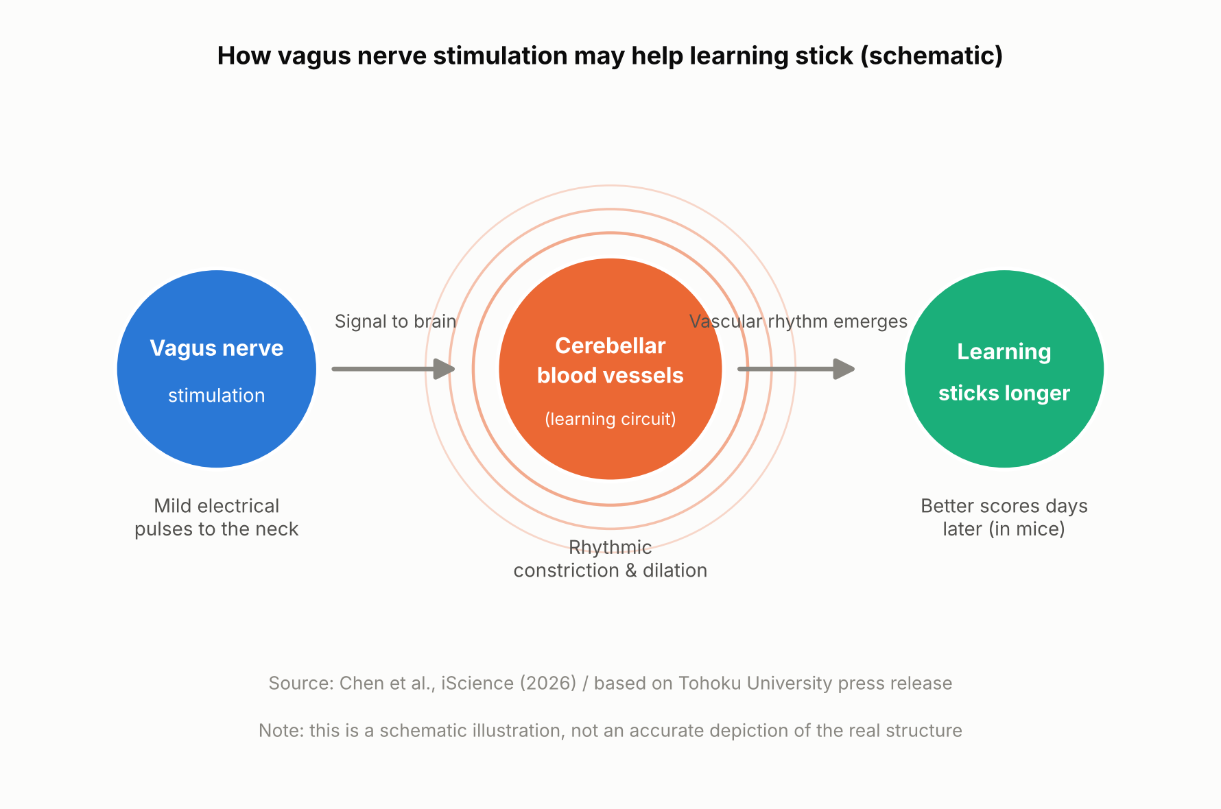
- Tohoku University researchers stimulated the vagus nerve while tracking local blood-volume changes in the brain, and found that repeated stimulation produced rhythmic fluctuations in local blood volume.
- Mice with larger blood-volume oscillations after stimulation tended to score higher on a learning test five days later. This is a mouse study, though, and it’s not yet known whether the same effect would occur in humans.
The Vagus Nerve — a Key Line Between Brain and Body
The vagus nerve is a major nerve that runs from the brainstem through the neck, chest, and abdomen, carrying signals between the brain and organs such as the heart, lungs, and digestive tract. It’s one of the main nerves connecting the brain and internal organs in both directions: it carries signals about the body’s internal state — such as heart rate, breathing, and digestion — up to the brain, while also carrying signals from the brain back down to those organs.
A treatment that stimulates this nerve from the outside with electrical pulses is called “vagus nerve stimulation,” or VNS. Using an implanted device, it’s an established therapy for epilepsy that doesn’t respond well enough to medication. In recent years, researchers have also been studying a non-invasive approach that stimulates part of the ear through the skin — though that’s a different method from the one used in this mouse study. Exactly what VNS changes inside the brain has remained unclear.
The Mouse Experiment: Stimulation Came After Practice
A team at Tohoku University’s Graduate School of Life Sciences — Junyu U. Chen, Assistant Professor Yoko Ikoma, and Professor Ko Matsui — fitted a small cuff electrode around the left cervical vagus nerve in mice and delivered electrical stimulation. The mice practiced a task called the horizontal optokinetic response (HOKR): tracking a vertical striped pattern that moved back and forth in front of them with their eyes. This kind of task is known to depend heavily on the cerebellum, a brain region central to motor learning.
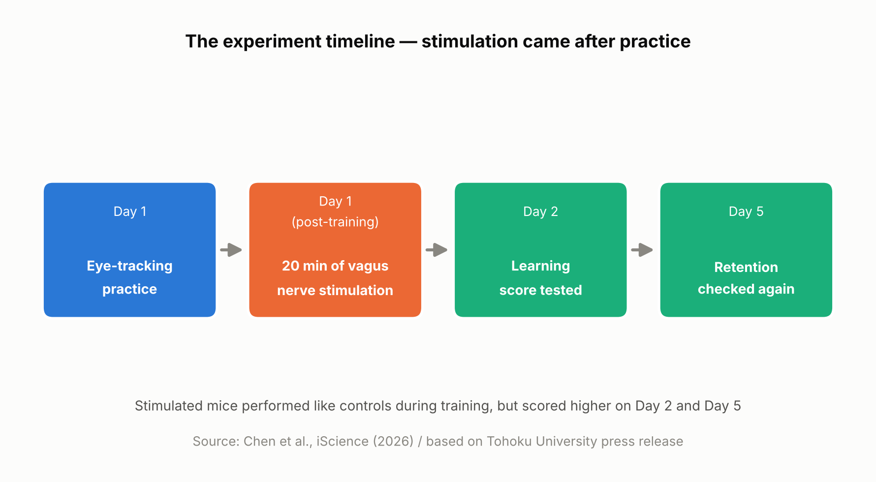
On day one, the mice practiced for 15 minutes, four times, spaced an hour apart. Right after each day-one training session, mice in the stimulated group received 20 minutes of vagus nerve stimulation at one of two intensities, while control mice received none. Performance was then measured again on day two and day five.
On day one itself, there was no difference between the stimulated and unstimulated mice. But by day two and especially day five, the stimulated mice showed higher scores, with the higher-intensity stimulation producing a visible benefit as early as day two and both intensities showing an advantage by day five. In other words, VNS didn’t directly boost performance during training — it appears to have improved later-day learning through some process that unfolded after training ended.
Blood Volume Around the Cerebellum Fell Into Rhythm After Stimulation
Using a light-based technique called fiber photometry, the researchers measured local blood-volume changes in tissue around the cerebellar flocculus. Each train of vagus nerve stimulation produced a two-step change: local blood volume briefly dipped, then — after a short delay — rose. This pattern is consistent with alternating contraction and dilation of blood vessels in that area.
With repeated stimulation, this rise-and-fall pattern settled into a steady rhythm. The researchers think this rhythmic change in blood volume may be related to how the brain regulates local blood flow and its metabolic environment — but the study did not directly measure oxygen, ATP, or other metabolic markers, so exactly how the blood-volume rhythm might support learning is still unclear.
Long-term learning is thought to involve various changes in the brain, including “synaptic plasticity” — changes in the connections and activity between neurons — which requires energy. The research team’s idea is that if the blood-volume rhythm is tied to how well-nourished brain tissue is, it could be helping support those changes, though this remains a hypothesis rather than something the study directly confirmed.
Mice with larger blood-volume oscillations after stimulation tended to score higher on the learning test five days later
A Bigger Rhythm, Better Long-Term Learning — but Only a Correlation So Far
The most notable finding was a relationship between the magnitude of the blood-volume oscillations in the 20 minutes right after stimulation and performance five days later: mice with larger oscillations tended to score higher on day five. It’s worth being clear that this is a correlation, not proof that the blood-volume change caused the better learning.
“The key point is that VNS was delivered only after training,” said Professor Matsui. “Our findings suggest that VNS may open a hidden window of opportunity for enhanced learning by making the brain environment more receptive to long-lasting change.” Lead author Junyu Chen added, “Our brains may be more strongly influenced by the body than we imagine. By tuning the brain’s metabolic environment, including rhythmic vascular movements, we may eventually unlock capacities that would otherwise remain latent.”
What’s Still Unknown — and What Comes Next
Based on this study alone, it’s not possible to say whether the blood-volume rhythm actually causes the learning improvement. Confirming that would require future experiments that directly manipulate blood-vessel or blood-volume activity and track the effect on learning. The researchers also did not directly measure metabolic molecules such as ATP, so the proposed mechanism remains a hypothesis.
It’s also worth remembering that this was a mouse study using an invasive cuff electrode surgically placed around the cervical vagus nerve, delivering stimulation directly to the nerve rather than through the skin. Less invasive, ear-based stimulation methods are already being studied in humans for other purposes, but whether this mouse study’s results would carry over to humans — or to those non-invasive methods — can’t be determined from this result alone. The research team suggests that fine-tuning the timing and intensity of stimulation could eventually help support learning and rehabilitation, though that application remains a research goal rather than something available today.
Sources: Tohoku University Graduate School of Life Sciences press release (August 26, 2026), Chen et al., iScience (published online August 25, 2026)
Leave a Reply