Category: Science

  • [Science] A Pufferfish’s Umami Sensor Is Broader Than Ours — What Its Structure Reveals About Why Animals Taste the World Differently

    [Science] A Pufferfish’s Umami Sensor Is Broader Than Ours — What Its Structure Reveals About Why Animals Taste the World Differently

    • Different animals can end up finding very different things “delicious” — and new research shows part of the reason comes down to the exact molecular shape of their taste sensors.
    • A Japanese research team used the SPring-8 synchrotron facility to determine the structure of the ligand-binding region of an umami receptor from pufferfish — the part that recognizes taste molecules.
    • The human umami receptor is highly selective: it responds strongly to only one mirror-image form of its target amino acid, and barely at all to the other. The pufferfish receptor, the team found, responds to both.
    • This is so far a test-tube finding about how the receptor binds molecules, not direct proof of what a pufferfish actually tastes. The difference may be related to the pufferfish’s diet, and the discovery could eventually help with new seasonings or aquaculture feed.

    Why the Same Food Doesn’t Taste the Same to Every Animal

    Umami — alongside sweet, sour, salty, and bitter — is one of the five basic tastes. It’s the savory taste produced when a receptor on the tongue detects certain amino acids, most notably glutamate, the compound behind the savory taste of kombu seaweed and dried bonito flakes. A receptor is a protein that changes shape when a specific molecule binds to it, and that shape change is what sends a signal into the cell. Scientists have long pictured this as a “lock and key”: a molecule has to be the right shape to fit and trigger a response.

    It has been difficult to determine exactly why umami receptors differ between species, partly because their three-dimensional structures are technically difficult to solve. Until now, structures of these receptors had been determined for only a small number of species, leaving the molecular basis of these differences unclear — even though different animals clearly do not all find the same molecules appetizing.

    Capturing the Pufferfish Receptor’s Shape at SPring-8

    A research team led by Professor Atsuko Yamashita at the Institute for Protein Research, Osaka University, working with Meiji University, Okayama University, Science Tokyo, and RIKEN, used X-ray crystallography to determine the crystal structure of the ligand-binding domain of the Tas1r1/Tas1r3 receptor complex — the extracellular region where taste molecules bind — in the tiger pufferfish (Takifugu rubripes, known in Japan as torafugu).

    The work was carried out at SPring-8, a large synchrotron radiation facility in Hyogo Prefecture, Japan. By aiming X-rays at extremely small protein crystals and analyzing the resulting diffraction pattern, the team worked out the three-dimensional structure of the pufferfish receptor’s taste-binding region for the first time.

    A Receptor That Also Responds to the “Mirror Image”

    Amino acids found in nature can exist in two mirror-image forms, often described loosely as “left-handed” and “right-handed” — what chemists call L- and D-amino acids. The human umami receptor strongly recognizes only the left-handed form — the same form as L-glutamate, the umami compound in kombu — and responds only weakly, if at all, to the right-handed form. To humans, that right-handed form often tastes sweet rather than savory.

    The new study found that the pufferfish receptor binds, and sends a signal in response to, both forms. Measuring binding strength directly, the team found the receptor bound the left-handed form of the amino acid alanine strongly, at a concentration of about 0.126 millimolar, while it bound the right-handed form more weakly, at about 1.44 millimolar — roughly a tenfold difference. Weaker binding still triggered the receptor, though. It’s worth being precise about what this does and doesn’t show: the result demonstrates that the isolated receptor binds and signals in response to the right-handed molecule in a test tube. It does not, on its own, prove that a live pufferfish actually perceives that molecule as delicious.
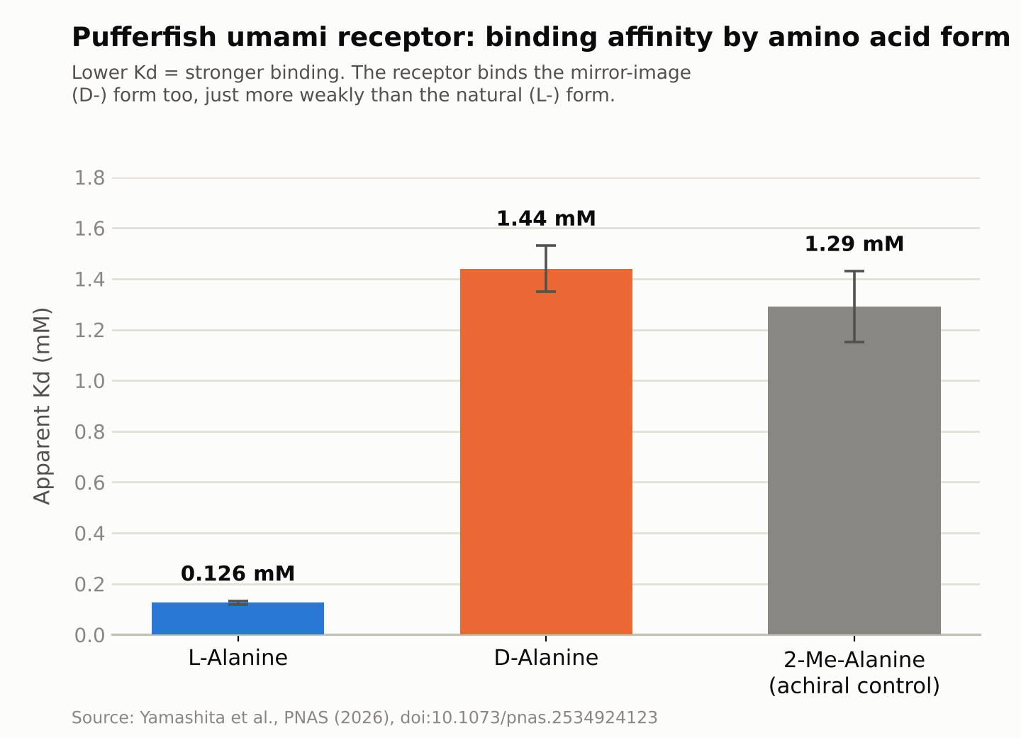
    Bar chart comparing how strongly the pufferfish umami receptor binds left-handed versus right-handed amino acids
    Source: created from data in Yamashita et al., PNAS (2026). A lower value means stronger binding.

    A Molecular “Latch” That Holds the Receptor Shut

    The receptor resembles a two-lobed clamshell that closes around a bound molecule of the right shape, and that closing motion is what sends the taste signal. The right-handed amino acid doesn’t fit neatly into the human version of this receptor. When the team examined the pufferfish receptor’s structure closely, they found an additional set of interactions between the two lobes — acting like a molecular latch — that helps keep the binding cleft closed even when the bound molecule’s chirality doesn’t match the usual, better-fitting form. That latch appears to let the receptor keep signaling even while holding a right-handed amino acid that would otherwise bind too loosely to trigger a response.

    The researchers suggest that this broader receptor specificity may be related to the pufferfish’s diet: mollusks and crustaceans, which make up much of what it eats, contain relatively high amounts of right-handed (D-) amino acids. Whether pufferfish actually perceive these molecules as tasty in the way we understand taste is a separate question the receptor data alone cannot answer — but the researchers note it as a plausible evolutionary explanation for why the receptor tolerates both forms.

    Toward New Seasonings and Aquaculture Feed

    What the study shows is limited to the structure and binding behavior of an isolated receptor studied in the lab; it doesn’t directly confirm how a living pufferfish experiences taste. Still, the research institutions involved say the structural information could eventually be useful for designing new umami compounds suited to the human palate, or for developing feed that farmed fish and livestock find more palatable — a question worth attention in aquaculture, where improving feed acceptance can matter for production efficiency and cost. The team also says it hopes to compare receptor shapes across more species going forward, providing structural information that could eventually help develop AI models for predicting how taste receptors recognize different molecules.

    Sources: Meiji University press release, RIKEN press release, Mizoguchi, Toda, et al., PNAS (2026)
  • [Science] APOE4 May Drive Scarring of Brain Blood Vessels — and a Human Brain-Tissue Model Showed the Process Can Be Reversed

    [Science] APOE4 May Drive Scarring of Brain Blood Vessels — and a Human Brain-Tissue Model Showed the Process Can Be Reversed

    • About one in four people carry a common gene variant that raises their odds of developing Alzheimer’s disease later in life — and scientists have now pinpointed, for the first time, exactly what that variant does to blood vessels in the brain.
    • The variant, called APOE4, is something people are simply born with; it isn’t caused by diet, exercise, or lifestyle.
    • Researchers found that APOE4 causes the cells that protect brain blood vessels to turn into “scar-forming” cells, making those vessels stiffer and more likely to clog.
    • The same team then found a way to brake that process in lab experiments, and even partly undo it.
    • The work is still limited to lab-grown tissue and animal models, but it points to a future where carrying this gene variant may not simply be something people have to live with — a treatment that slows or blocks the damage could eventually become possible.

    A Gene Long Blamed, Now Tied to a Specific Mechanism

    Scientists have linked APOE4 to Alzheimer’s since landmark studies in 1993, and subsequent research has implicated it in several disease-related processes, including amyloid-β metabolism and clearance, lipid handling, and inflammation. What has stayed less clear is exactly how APOE4 damages brain blood vessels, an abnormality long observed in carriers alongside the disease’s hallmark protein buildup. In a paper published in the journal Cell on September 24, 2026, a team led by Dr. Joel W. Blanchard at Mount Sinai pinned the vascular damage to a specific cell-identity switch, using evidence from human brain tissue, lab-grown human brain models, and mice.

    How a Support Cell Turns Into Scar Tissue

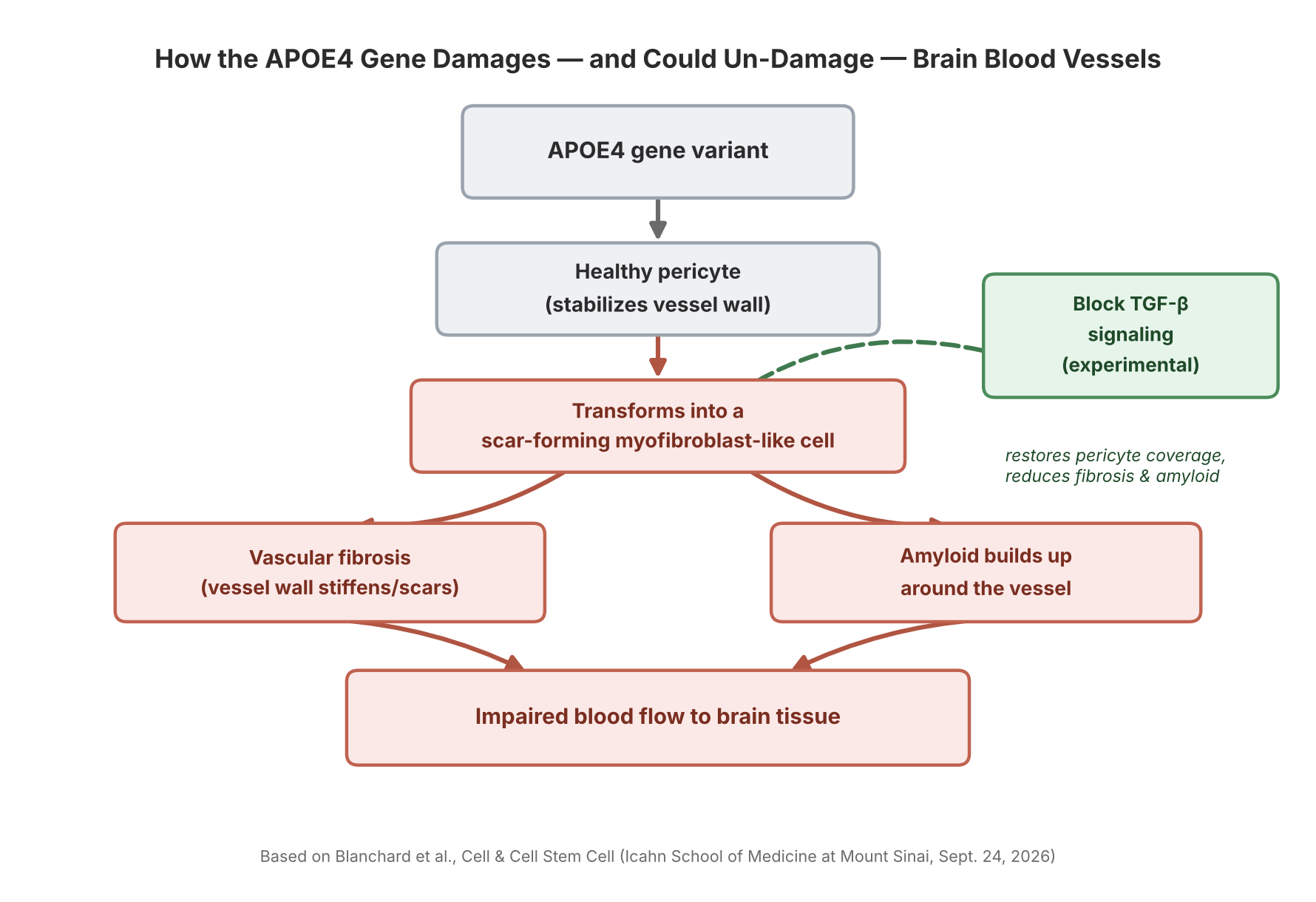
    Tiny blood vessels in the brain are wrapped in pericytes, cells that help stabilize the vessel wall and maintain the blood-brain barrier. The Mount Sinai team found that APOE4 pushes a portion of these pericytes into a myofibroblast-like state — a cell identity similar to the scar-forming cells involved in fibrosis in organs such as the liver and lungs. As more pericytes shift into this state, they produce a dense, fibronectin-rich extracellular matrix that accumulates in the vessel wall (a process called vascular fibrosis) and promotes the buildup of amyloid protein around the vessel. Mount Sinai’s own description of the work says these changes “may compromise blood flow” and contribute to neurodegeneration — a more cautious framing than saying blood flow is cut off outright, since that specific chain of cause and effect in people has not been directly demonstrated.

    Diagram: how APOE4 damages and could un-damage brain blood vessels
    Source: based on Blanchard et al., Cell & Cell Stem Cell (Icahn School of Medicine at Mount Sinai, Sept. 24, 2026)

    Built From Human Brain Tissue, Not Just Mice

    A notable part of this work is how closely it was tied to human biology. The researchers assembled a single-cell transcriptomic atlas of the human brain’s blood vessels, then examined the pericyte-to-myofibroblast switch in miBrains — a lab-grown, three-dimensional tissue model derived from human induced pluripotent stem cells that contains multiple brain cell types, including vascular cells — as well as in postmortem human brain tissue. They also examined the mechanism in aged APOE4 mice. The clearest demonstration that the process could be reversed came from the miBrain model: blocking TGF-β signaling there restored pericyte coverage and reduced fibrosis and vascular amyloid. Combining human tissue data with a human-cell model and an animal model, rather than relying on mice alone, gives the mechanism more weight as a plausible driver of disease in people — though it is still evidence from models, not a demonstration in living human patients.

    A Second, Related Finding: Cholesterol and the Brain’s Cleanup System

    A companion paper from the same group, published online in Cell Stem Cell on August 24, 2026, points to a related problem in a different brain cell type. The team found that APOE4 causes cholesterol to accumulate inside astrocytes — star-shaped support cells involved in clearing cellular waste through the endolysosomal system, the cell’s internal recycling and degradation pathway. That cholesterol buildup impaired the astrocytes’ ability to process alpha-synuclein, the protein that forms the toxic clumps central to Parkinson’s disease and Lewy body dementia, and which can also appear as a secondary pathology in some Alzheimer’s cases. Together, the two papers point to two distinct cellular mechanisms through which APOE4 may contribute to neurodegenerative disease, though the papers don’t claim these are the only routes involved.

    Blocking TGF-β signaling “restored pericyte coverage and reduced both fibrosis and vascular amyloid” in the researchers’ experimental models — evidence that at least some APOE4-associated vascular abnormalities are not necessarily permanent.

    Why “Reversible in Models” Is the Real Headline

    The most consequential detail in the Mount Sinai findings isn’t the damage itself — it’s that part of it could be undone experimentally. The pericyte-to-myofibroblast switch is driven by a signaling pathway called TGF-β. When the researchers blocked TGF-β signaling in their models, pericytes stopped converting into scar-forming cells, and both vascular fibrosis and amyloid buildup around the vessels were reduced. That gives researchers a specific, experimentally testable target to study further: TGF-β signaling, along with the lipid-metabolism and endolysosomal pathways highlighted in the companion paper, now stand out as candidate starting points for future research into Alzheimer’s and other neurodegenerative diseases involving alpha-synuclein pathology — a different starting point than simply trying to clear amyloid after it has already built up.

    What This Means If You Carry the Gene

    None of this is a treatment, and the reversal was shown in cell and animal models, not in people — any drug built on these targets would still need years of testing before reaching patients. But the finding reframes what APOE4 does: carrying the variant doesn’t guarantee disease, and it isn’t an on/off switch for Alzheimer’s — rather, it can shift several biological processes, including this pericyte transition, in ways that raise vulnerability. For the roughly one in four people who carry an APOE4 copy, the significance of this research is that it offers a starting point for seeing APOE4 not only as a risk marker, but as a set of specific cellular mechanisms that future research could study, and potentially target, directly.

    Sources: Mount Sinai Newsroom, Cell, Cell Stem Cell, National Institute on Aging

  • [Science] Swiss Glaciers Lost 5.5% of Their Ice in 2026 — A Preview of Tighter Summer Water Supplies Across the Alps

    [Science] Swiss Glaciers Lost 5.5% of Their Ice in 2026 — A Preview of Tighter Summer Water Supplies Across the Alps

    Switzerland’s glaciers lost about 5.5% of their ice volume during the 2025/2026 measurement year, the second-largest annual loss in the GLAMOS monitoring record after 2022. Over the past five years, Switzerland has lost nearly one-fifth of its glacier ice — a five-year loss rate more than twice that of any previous five-year period in the record. The pace matters well beyond Switzerland’s borders: meltwater from its glaciers helps sustain the Rhône, the Rhine, and other rivers that millions of people downstream rely on for drinking water, farming, and electricity, particularly during hot, dry summers.

    A second-worst year, right after the worst five-year stretch on record

    The figures come from GLAMOS, Switzerland’s long-term glacier monitoring network, run jointly by ETH Zurich and the universities of Fribourg and Zurich, which carries out detailed annual mass-balance measurements on 23 Swiss glaciers each year. Its 2025/2026 annual report puts the average “mass balance” — the net change in a glacier’s mass, expressed as meters of water equivalent (m w.e.), a way of converting accumulated snow minus melt into an equivalent depth of water spread over the glacier — at −2.86 m w.e. That is the second-most negative year GLAMOS has recorded, behind only 2022’s −3.13 m w.e.

    Converted into ice volume, the 2025/2026 melt amounted to roughly 5.5% of Switzerland’s glacier ice. Ten of the 23 monitored glaciers lost more than 3 meters of water equivalent; Tsanfleuron fared worst at −3.9 m w.e., while Findel, near the Matterhorn, recorded the smallest loss, at −2.3 m w.e. Four glaciers — Allalin, Clariden, the Grosser Aletsch (the largest glacier in the Alps), and the Rhone glacier — recorded the greatest annual mass loss in their observational history.

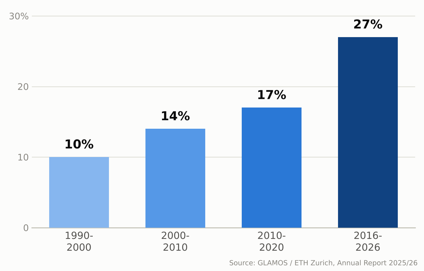
    Zoom out and the five-year picture is starker. Since 2021, Swiss glaciers have lost close to 20% of their remaining volume — a loss rate GLAMOS describes as more than twice as fast as any earlier five-year period in its record. The longer-term picture shows the same acceleration: Swiss glaciers lost about 10% of their volume in 1990–2000, 14% in 2000–2010, 17% in 2010–2020, and about 27% over the most recent ten-year window, 2016–2026.

    Bar chart showing Swiss glacier ice volume loss by decade: 10% in the 1990s, 14% in the 2000s, 17% in the 2010s, and 27% in 2016-2026
    Share of ice volume lost by Swiss glaciers, by decade. Source: GLAMOS / ETH Zurich, Annual Report 2025/26.

    Why this past year was so bad

    Two seasons combined to make 2025/2026 so damaging. Winter snowfall came in about 25% below the long-term average by the end of April, leaving glaciers with a thin protective layer heading into summer. The following April–September period was, according to glaciologist Matthias Huss, who leads GLAMOS and is a professor at ETH Zurich, the hottest and driest “summer half-year” in Switzerland since national weather records began in 1864. The freezing level — the altitude of the 0°C mark in the atmosphere — rose above 4,000 meters on 76 days, more than double the average and a new Swiss record. By September, snow cover had disappeared even at elevations around 3,500 meters.

    GLAMOS calls the point when melt overtakes the winter’s snow gains — after which every melted liter comes straight out of the glacier’s mass — “Glacier Loss Day.” In 2026 that point arrived on June 29, the second-earliest date on record, three days later than the record set in 2022. One reason 2026 still fell just short of 2022’s record was the smaller amount of Saharan dust deposited on the snow that year, which let glaciers reflect more sunlight than they did in 2022, when dust darkened the surface and accelerated melting.

    Melting is also carving cavities and collapse zones into some glaciers

    Researchers monitoring the retreat describe an additional process layering on top of ordinary surface melting. On some glacier tongues, meltwater penetrates and flows through the ice, forming channels and cavities; where these grow large enough, sections of ice can collapse inward rather than simply retreating at the edges. Huss has described visiting high-altitude sites that were still covered in ice a few years ago — including areas near the Grosser Aletsch’s Konkordiaplatz and around the Findel glacier by the Matterhorn — and finding bare, rubble-strewn rock he compared to a lunar landscape.

    Some glaciers are also disappearing outright. The Bella Tola glacier in canton Valais, which had been measured for decades, is among the small glaciers researchers now consider completely and permanently gone as of 2026. GLAMOS estimates that roughly another 100 small Swiss glaciers disappeared between 2016 and 2023 — a pace this year’s melt appears to be extending rather than slowing.

    A record summer of meltwater is also a warning sign

    Between July and September 2026 alone, Swiss glaciers released an estimated 2.2 trillion liters of water — more than four times the amount all Swiss households drink in a year.

    Between July and September 2026, Swiss glaciers released roughly 2.2 trillion liters of meltwater — equivalent to more than four times the annual drinking-water consumption of every household in Switzerland.

    That sounds like abundance, and in the short term it was: the extra runoff topped up rivers during a hot, dry summer when demand for water was high. But glaciologists are blunt about what it really represents — a one-time withdrawal from a shrinking reserve, not a renewable supply. Glaciers act as a buffer that releases extra meltwater precisely in hot, dry years when it’s needed most; snowfall can replenish that mass over time, but only when accumulation exceeds losses, which has not been the case in most recent years. As the ice reserve keeps shrinking, its ability to smooth out future droughts shrinks with it.

    That matters beyond Switzerland too. Glacier melt contributes to summer runoff in Alpine river basins, including the Rhône and the Rhine, helping sustain flows during hot, dry periods for communities downstream in France, Germany, and beyond. The 2026 season offered unusually generous meltwater precisely because the ice had so much to give up. A smaller, thinner body of Alpine glaciers will have less stored ice to release in the next hot summer, and some smaller glaciers may disappear entirely.

    What the long record shows

    GLAMOS’s value lies partly in its length: continuous measurements stretching back decades let scientists tell a single rough year apart from a structural shift, and the longer-term record shows increasingly rapid losses, with the most recent ten-year period (2016–2026) substantially exceeding the losses seen in each earlier ten-year period. By the end of the 2025/2026 measurement season, Switzerland’s glaciers held an estimated 42.6 cubic kilometers of ice across 714 square kilometers of glacier-covered area — down by more than 30 cubic kilometers, and about a third in glacier-covered area, since 2000.

    Researchers caution against reading any single year, including 2026’s near-record losses, as proof of exactly what comes next; Alpine weather still varies year to year, and a cold, snowy winter can briefly slow the trend. But the multi-decade pattern GLAMOS has documented — each ten-year period worse than the last — is increasingly relevant to how water-resource managers, hydropower operators, and mountain communities across the Alps plan around the region’s future water supply.

    Sources: ETH Zurich, “Devastating year for Switzerland’s glaciers”, GLAMOS, Annual Report on Swiss Glaciers 2025/26, PBS NewsHour / Associated Press

  • [Science] The Instant You Decide, Your Brain Re-Files the Options — MIT Neuroscientists Find a Flexible Code That Could Inform AI Design

    [Science] The Instant You Decide, Your Brain Re-Files the Options — MIT Neuroscientists Find a Flexible Code That Could Inform AI Design

    Every time you choose between two options — which job offer to accept, which product to buy — your brain does more than simply retrieve what each one was worth. A new MIT study shows that the instant you make that choice, the neural code representing those options in part of your brain reorganizes itself, shifting from grouping information by the order you saw it to grouping it by whether you picked it. The finding, published online in the journal iScience in September 2026, offers a rare, cell-by-cell look at how flexible biological memory can be — and it gives researchers working on adaptive AI a biological case study worth studying, even though the experiment itself did not involve any AI system.

    Why Scientists Wanted to Watch the Brain Mid-Decision

    Researchers have long known that the prefrontal cortex, the region of the brain just behind the forehead, plays a central role in weighing options and making choices. Many of its neurons also show “mixed selectivity,” meaning a single cell’s activity can shift depending on a combination of task variables rather than tracking one fixed thing. What earlier work left unresolved was how the brain avoids mixing up two options that were evaluated only moments apart, especially once one has been chosen and the other set aside. A team led by MIT graduate student Huidi Li and senior author Earl K. Miller, a professor at MIT’s Picower Institute for Learning and Memory, set out to track that process neuron by neuron rather than assume an answer. The team, which also included Nikolaos Chrysanthidis, Scott Brincat, and Jonas Rose, published its results in iScience; MIT publicized the findings on September 22.

    Watching Hundreds of Neurons Decide, in Real Time

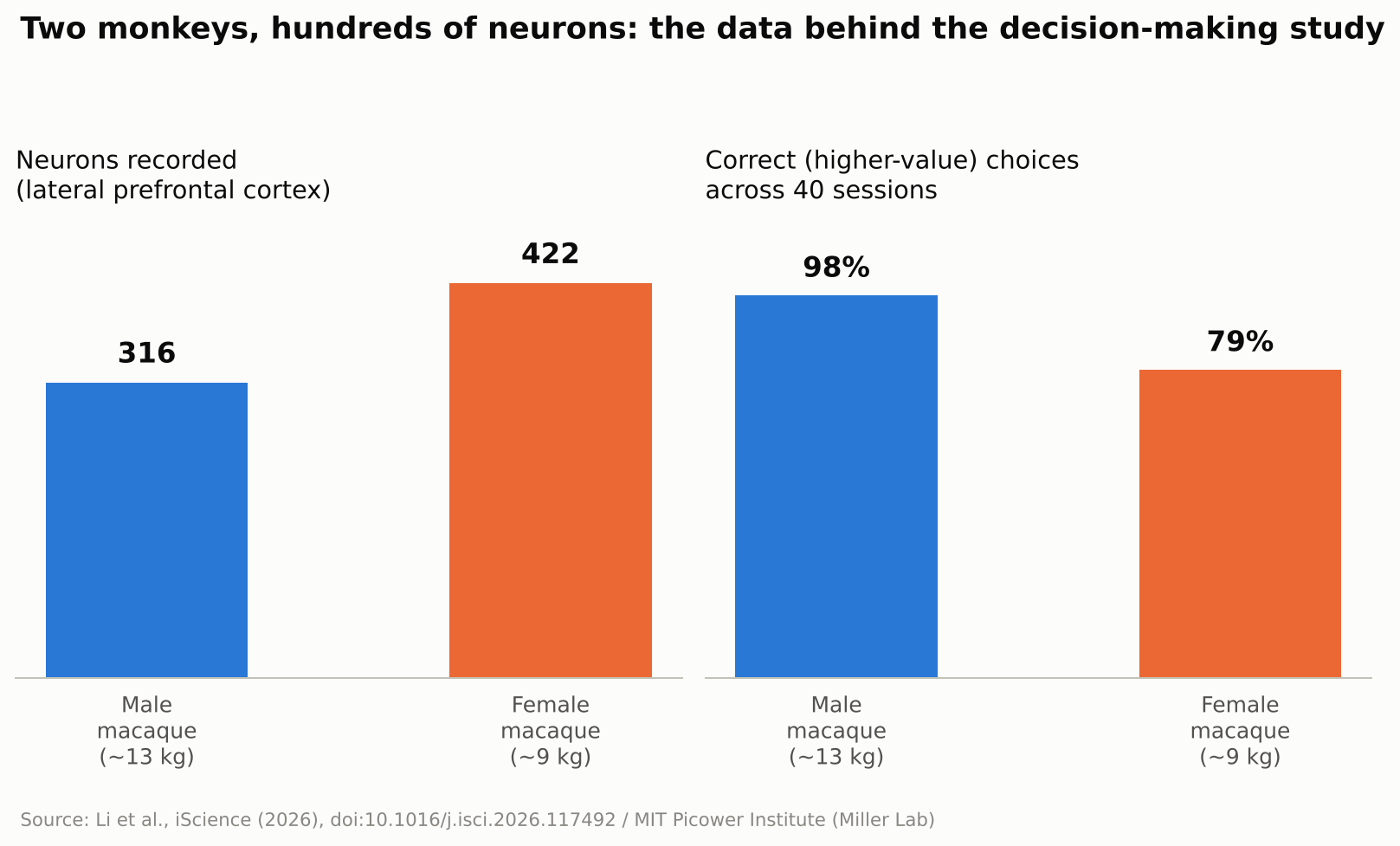
    To find out, the researchers trained two macaque monkeys — a roughly 13-kilogram male and a 9-kilogram female — to play a simple reward game. A white square flashed briefly at one of four spots on a screen, followed by a colored ring that signaled how many drops of juice that option was worth, anywhere from one to thirteen. The same sequence then repeated for a second target at a different screen location. Once both options had been shown, the monkey looked toward its preferred target and received the corresponding juice reward. The male selected the higher-value option on about 98 percent of trials; the female, on about 79 percent. While the animals made their choices, the team recorded the electrical activity of 316 neurons in the male’s lateral prefrontal cortex — the side portion of the prefrontal cortex — and 422 in the female’s, across 40 recording sessions. Rather than tracking single cells in isolation, the researchers analyzed the activity of the whole recorded population as points moving through a high-dimensional mathematical space, a technique known as neural subspace analysis: when enough neurons fire in a similar combined pattern, that pattern occupies its own region, or “subspace,” within that space, and two subspaces that barely overlap are described as close to orthogonal, or mathematically independent of each other.

    Bar chart showing neurons recorded and choice accuracy for two macaques in the study
    Source: Created from data in Li et al., iScience (2026)

    Before the Choice: Order. After the Choice: Outcome.

    Seen through that lens, the result is a clear shift in organization. Before a choice was made, the subspaces representing the first-shown and second-shown targets were kept largely separate, organized around presentation order. Once the monkey chose, the picture changed: the subspaces reorganized around the decision itself, with the chosen and unchosen options rotating into distinct, close-to-orthogonal subspaces regardless of the order in which they had originally appeared. In other words, the brain’s filing system for “what I saw first and second” gave way to a new one built around “what I picked and what I didn’t.” The researchers suggest this reorganization helps downstream brain regions read out the decision efficiently, since they could rely on a consistent format rather than track which position each option had occupied.

    “The brain doesn’t just hold information statically,” Li said.

    No Single Neuron “Owns” a Decision

    A second notable result was how that reorganization happened at the level of individual cells. The researchers found no fixed set of neurons permanently dedicated to representing “the chosen option” or “the rejected one.” Instead, individual neurons shifted which ensemble they contributed to from one trial to the next, joining a different, temporary coalition of cells depending on what the task required at that moment. Researchers in the field generally see this kind of flexible, overlapping coding as a way for a limited number of neurons to represent many different combinations of information, rather than requiring a dedicated cell type for every possible scenario — though this study did not directly measure how much representational capacity that flexibility adds.

    Why This Matters Beyond the Lab

    The study did not test any artificial intelligence system, and the researchers were not trying to build one. Still, the result speaks to a question AI researchers care about: how can a learning system keep updating what it represents without losing track of information it already holds? Many AI systems, once trained, represent what they have learned in ways that stay largely fixed afterward, which is part of why training them further on new material can degrade old knowledge, a problem researchers call catastrophic forgetting. The MIT findings do not explain or solve that problem, but the underlying idea — a biological circuit reorganizing its internal representations on the fly to match what a task demands at each stage — is the kind of mechanism that researchers in continual learning and brain-inspired, or neuromorphic, computing study for design ideas. It is also a small, concrete answer to a more everyday question: the researchers found that even after a choice was made, the unchosen option remained represented in its own distinct subspace rather than disappearing from the recorded activity. MIT’s own description of the work suggested this could be important for later crediting an outcome to the option that produced it, which may help the brain learn from an alternative it did not select — though that specific learning process was not directly tested in this experiment.

    What’s Still Unknown

    The authors are careful to flag the limits of the work. Only two animals took part, and the task involved a simple, sequential choice between two options with clearly different, discrete reward values — closer to comparing two job offers one after the other than to scanning a crowded menu of possibilities at once. Whether the same kind of reorganization occurs when values vary continuously, when there are more than two options on the table, or in human decision-making specifically, remains an open question the team says future studies will need to address. Macaque and human prefrontal cortex share many organizational similarities, which is why monkey studies like this one are routinely used to generate hypotheses about human cognition, but confirming that the same reorganization happens in people would require a separate study in humans. It is also worth remembering that this was a recording study: it shows that representations change alongside decisions, not that the reorganization itself is what causes a choice to happen. As with any single study, independent replication will matter before the finding is considered settled.

    For now, the study offers a concrete, carefully measured answer to a question that sounds almost philosophical: what happens in your head in the instant after you decide? According to this research, at least in one brain region of two monkeys performing a simple task, the answer is that the brain quietly rebuilds how it was organizing two competing options around the choice that was just made.

    Sources: MIT News, iScience (Li et al., 2026), EurekAlert!

  • [Science] The Ice Shooting Off Saturn’s Moon Enceladus Isn’t a Simple Copy of Its Ocean — A Finding That Could Reshape the Hunt for Alien Life

    Every second, the icy moon Enceladus sprays countless grains of ice from its hidden ocean into space, feeding one of Saturn’s rings. An international team led by the Freie Universität Berlin and the Earth-Life Science Institute at the Institute of Science Tokyo has proposed that, on the way out, that water gets chemically sorted. The upshot: an individual ice grain is not simply a miniature sample of the whole ocean, which matters for any future mission trying to read Enceladus’s ocean chemistry for signs of life.

    Original source
    Journal: Science Advances (DOI: 10.1126/sciadv.aee7256)
    Press release: Freie Universität Berlin
    Data: 961 ice-grain spectra recorded by Cassini’s Cosmic Dust Analyzer

    Cooling speed decides which salt goes where

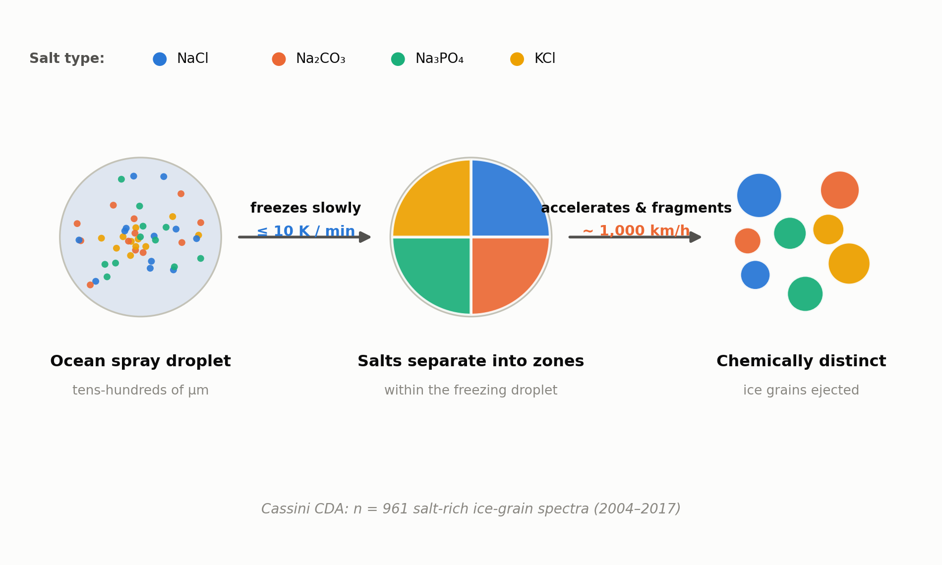
    The team ran lab experiments with droplets formulated to match the salts expected in Enceladus’s subsurface ocean. When droplets roughly 200 micrometers across were cooled slowly, at about 10 kelvin per minute or less, the dissolved salts did not freeze evenly into the ice. Instead, compounds such as sodium chloride, carbonates, phosphates and potassium salts separated out and concentrated into distinct regions within the droplet as it froze. Rapid freezing did not produce the same pronounced separation, the experiments showed.

    961 oddly different ice grains from Cassini

    What happens to a droplet after it freezes unevenly underground? The team proposes that as these droplets rise through narrow vent channels toward the surface, they accelerate — reaching roughly 1,000 km/h in the model — before shattering against the icy channel walls into the fine grains that eventually reach space. NASA’s Cassini spacecraft, which orbited Saturn from 2004 to 2017, carried a Cosmic Dust Analyzer (CDA) that recorded mass spectra for 961 salt-rich ice grains gathered over the course of its mission. Grain to grain, the chemistry varied widely — some grains dominated by sodium salts, others by carbonates or phosphates — even though they presumably all came from the same ocean. The new freeze-and-fragment model offers an explanation for why that is: each grain simply froze out of a different, already-segregated pocket of the original droplet.

    “The oceanic constituents are separated from each other and simultaneously concentrated into individual ice particles,” says Professor Frank Postberg of Freie Universität Berlin.
    Schematic of the proposed ice-grain formation model at Enceladus
    Source: created from data released by Freie Universität Berlin / Institute of Science Tokyo (proposed grain-formation model)

    A discovery that could change how we read the data

    The core implication is that no single ice grain can be assumed to represent the ocean’s average composition. That matters for mission concepts such as NASA’s proposed Orbilander, which would collect and analyze Enceladus’s plume grains directly: researchers will likely need to account for grain-to-grain differences when interpreting what they find. The team suspects organic molecules and other life-relevant compounds could be concentrated in a similar way, though confirming that will require further study.

    Enceladus remains one of the solar system’s most closely watched candidates for hosting life. Understanding how each ice grain came to be — rather than treating it as a simple drop of seawater — looks set to become an unavoidable consideration for the missions that follow.

    Xsciens editorial note

    This study is a model that explains an existing dataset, not a new observation of Enceladus. Its central claim, that cooling speed determines which salts end up in which ice grains, is a proposal that fits the 961 Cassini spectra. It has not been confirmed by a new spacecraft measurement. The reading is attractive because it ties grain composition to a physical process, but it also means any future mission, such as the Orbilander concept, would need to test it directly before the interpretation of the data is changed.

    Key terms

    • Cosmic Dust Analyzer (CDA): the Cassini instrument that analyzed the composition of individual ice grains in flight.
    • Cooling rate: how fast a droplet loses heat; the study ties it to which salts crystallize in a grain.
    • Vent: a fissure on Enceladus’ south pole through which water-rich material is ejected into space.

    Sources: Freie Universität Berlin press release, Science Advances (DOI: 10.1126/sciadv.aee7256)

  • [Science] A Day Isn’t Exactly 24 Hours — A Tug-of-War at Earth’s Core Is Behind the Multi-Decade Drift

    Every day you live through is not quite the same length as the last one, and the difference is measured in milliseconds. Part of this wobble, especially its slow, multi-decade rhythm, is tied to how a hidden solid ball of iron at Earth’s center, the inner core, pulls against the mantle above it, according to a new model-based analysis by researchers at the University of Alberta. The precise tracking of Earth’s rotation that underlies GPS navigation and the world’s official timekeeping is not unrelated to this invisible tug-of-war deep inside the planet.

    原論文情報 / Original Paper
    Title: Gravitational torque drives multidecadal variations in length of day
    Authors: Zhang, H. and Dumberry, M.
    Journal: Nature 658, 119–124 (2026)
    URL: nature.com/articles/s41586-026-10999-2

    A Tug-of-War Inside the Planet

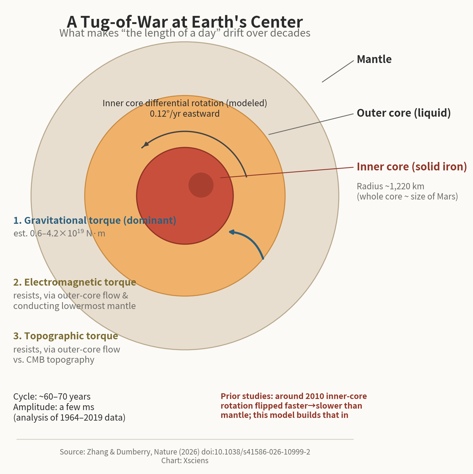
    Huifeng Zhang and Mathieu Dumberry, physicists at the University of Alberta in Canada, combined 55 years of Earth-rotation data (1964–2019) with a seismologically informed history of inner-core rotation and models of outer-core flow. They found that a roughly 60-to-70-year cycle in the length of day, with an amplitude of just a few milliseconds, can largely be explained by a model in which gravitational torque from the inner core is the main driver. Their paper appeared in the journal Nature on September 23, 2026.

    Roughly 5,000 km beneath the mantle sits a solid iron inner core with a radius of about 1,220 km, surrounded by the molten outer core (the inner and outer core together, as a single unit, are closer to the size of Mars). According to the researchers, a slight mismatch between the inner core’s shape and mass distribution and the denser regions of the mantle above generates what they call a gravitational torque, the main factor changing the rotation speeds of both the inner core and the rest of the planet.

    Diagram of the three torques (gravitational, electromagnetic, topographic) tugging on Earth's inner core
    Source: made from data in Zhang & Dumberry, Nature (2026)

    An Invisible Core, and How We Track Earth’s Spin

    Two other forces push back against that gravitational pull: a “topographic torque,” arising as outer-core flow interacts with bumps and dips on the core-mantle boundary (CMB), and an “electromagnetic torque,” transmitted through Earth’s magnetic field as the outer core flows past the electrically conducting lowermost mantle. Together they act as a brake. The model estimates the gravitational torque at roughly 0.6 to 4.2 × 10¹⁹ newton-meters, with the inner core creeping eastward relative to the mantle by about 0.12 degrees a year. None of these are direct measurements — they are values inferred from rotation data by working backward through the model.

    Prior seismological studies suggest that, around 2010, the inner core switched from rotating faster than the mantle to rotating slower — a pattern this new model builds into its calculations.

    These small shifts in Earth’s rotation matter for precisely tracking and predicting Earth’s orientation, which underlies satellite navigation, and for decisions about “leap seconds,” the adjustments occasionally used to keep Coordinated Universal Time aligned with Earth’s actual rotation. Earth’s spin has been trending slightly faster in recent years, and international timekeeping bodies are now discussing changes to how leap seconds are handled. Work like this offers a way to connect small, surface-level timing signals to physical models of a part of the planet no one can observe directly.

    Because no one can drill down to the inner core, scientists have had to infer its properties indirectly, mostly from how seismic waves travel through it. The authors themselves note that their results depend on the accuracy of the underlying models of inner-core rotation and outer-core flow. Still, connecting an everyday measure like the length of a day to physical models of Earth’s deep interior offers one more way to probe a part of the planet we may never see directly.

    Xsciens editorial note: inferred, not measured

    The key caution is in the article’s own wording: the torque strength (about 0.6 to 4.2 × 10¹⁹ newton-meters) and the inner core’s drift (0.12 degrees per year eastward) are values inferred through a model, not measured directly. They show that a gravitational-torque model can explain the observed 60-to-70-year cycle in the length of day, which is different from proving it is the only explanation.

    Another detail worth noticing is the time span. The rotation data cover 1964 to 2019, about 55 years, which is shorter than one full 60-to-70-year cycle, so the long rhythm is being inferred from less than a complete period. The model also builds in a seismology-based claim that the inner core switched around 2010 from turning faster than the mantle to turning slower. What to watch next is whether new seismic observations of the inner core and further years of rotation data keep agreeing with the model.

    Key terms

    • Inner core: the solid iron ball at Earth’s center, with a radius of about 1,220 km
    • Outer core: the liquid layer surrounding the inner core
    • Mantle: the thick rocky layer between the core and Earth’s crust
    • Torque: a twisting force that changes how fast something rotates
    • Core-mantle boundary (CMB): the interface between the outer core and the mantle
    • Leap second: an occasional one-second adjustment that keeps Coordinated Universal Time aligned with Earth’s rotation

    Sources: Zhang, H. & Dumberry, M. “Gravitational torque drives multidecadal variations in length of day.” Nature 658, 119–124 (2026), Nature News & Views commentary, ScienceDaily

  • [Science] Scientists Get Cancer Cells to Make Their Own Chemo Drug — Lab Tests Suggest a Way to Spare Healthy Tissue

    [Science] Scientists Get Cancer Cells to Make Their Own Chemo Drug — Lab Tests Suggest a Way to Spare Healthy Tissue

    Much of chemotherapy’s toll on the body comes from the fact that the drug attacks healthy, fast-dividing cells right along with cancerous ones. A team from RIKEN and Institute of Science Tokyo has developed a way around that: instead of administering an active drug, they deliver an inert precursor that only turns into a cancer-fighting compound once it is inside a cell, using carbon monoxide (CO) — a metabolite that runs elevated in some cancer cells — as the raw material. In effect, the cancer cell is enlisted to manufacture its own drug from a harmless precursor, while cells with lower CO output make far less of it. It’s an early-stage, cell-culture result rather than a ready-made treatment: the researchers themselves describe the selectivity they observed as modest, and animal or human safety data don’t yet exist.

    原論文情報 / Original Paper
    Title: Harnessing endogenous carbon monoxide for cancer-selective drug synthesis
    Authors: Kawai, M. et al.
    Journal: Nature Communications 17, 9603 (2026)
    DOI: 10.1038/s41467-026-77616-8

    A palladium catalyst smuggled in by the body’s own protein

    The team built what they call an albumin-palladium complex: a palladium catalyst tucked into a hydrophobic pocket of human serum albumin, the most abundant protein in blood. A small anchor molecule steers the complex to albumin’s drug-binding site, and this packaging keeps the catalyst active even in the presence of glutathione, an abundant cellular antioxidant that would normally deactivate a bare palladium compound. Once inside a cell, the complex reacts with intracellular CO to assemble a phenanthridinone-scaffold compound with topoisomerase-I-inhibiting activity — a mechanism shared with established anticancer drugs. The approach flips the usual order of drug delivery: rather than making a drug and shipping it to the cell, the team ships an inert precursor and lets the cell’s own CO serve as the building material for the final compound, assembled on site.

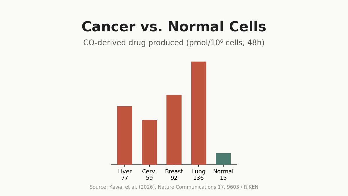
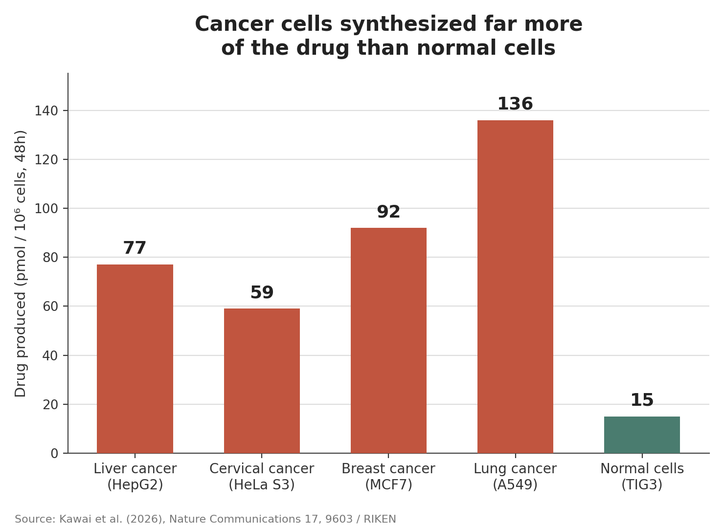
    Bar chart comparing CO-derived drug production across four cancer cell lines and one normal cell line
    Source: created from data in Kawai et al. (2026), Nature Communications 17, 9603

    Up to ninefold more compound formed in cancer cells

    According to the paper, after 48 hours in culture, the amount of CO-derived compound produced in four cancer cell lines — liver (HepG2), cervical (HeLa S3), breast (MCF7), and lung (A549) — was up to roughly ninefold higher than in normal fibroblasts (TIG3): 136 picomoles per million cells for the lung line versus 15 picomoles for the normal one. In the liver cancer line, the concentration needed to be effective (IC50) was 13.99 micromolar, about a third of the 37.54 micromolar needed against normal fibroblasts. In breast cancer cells, RIKEN’s release reports that treatment at 5 to 10 micromolar significantly suppressed colony formation over 12 days, with near-complete inhibition at 10 micromolar and above.

    After 48 hours, lung cancer cells used CO to synthesize roughly nine times as much of the drug compound as normal fibroblasts did.

    A new chemical angle on reducing chemotherapy’s toll

    The researchers frame this as an early step toward a broader drug-design strategy: using disease-specific metabolites, not just in cancer, as the raw material for synthesizing therapeutics inside the body rather than merely as diagnostic markers. The comparisons so far involve one cancer cell line and one normal cell line at a time, and the work is limited to cultured cells — animal and clinical studies still lie ahead. Still, it offers a new chemical approach to the long-standing challenge of designing cancer treatments that spare healthy tissue.

    The study was published on September 17, 2026, in Nature Communications.

    Xsciens editorial note: what the numbers do and do not show

    The most striking figure is the up to ninefold difference in how much compound the cells made, with lung cancer cells producing 136 picomoles against 15 picomoles in normal fibroblasts. But that is a measure of synthesis inside cells, not of tumor shrinkage. The effective concentration (IC50) in liver cancer cells, 13.99 micromolar versus 37.54 micromolar in normal fibroblasts, is about a third, so the selectivity in killing cells is real but more modest than the ninefold synthesis gap suggests.

    It is also worth noting how the idea differs from conventional targeted drugs. Those aim at a molecule on or in the cancer cell. Here, a harmless precursor is delivered first and the cancer cell’s own metabolism supplies carbon monoxide as a building block, so the strategy depends on how much CO a given tumor makes. The comparisons so far use single cell lines in culture, so how well this works in tumors made of mixed cells, and whether enough precursor can reach them, are open questions for animal and clinical studies.

    Key terms

    • Albumin: the most abundant protein in blood, here used as a carrier for the catalyst
    • Palladium complex: a compound of the metal palladium that acts as a catalyst, speeding up a chemical reaction
    • Glutathione: an antioxidant present in large amounts in cells that can deactivate many catalysts
    • Topoisomerase I inhibitor: a drug that blocks an enzyme cells need to copy DNA, a mechanism used by established anticancer drugs
    • IC50: the concentration of a compound needed to reduce cell growth by half

    Sources: RIKEN, Kawai et al., Nature Communications 17, 9603 (2026), Tech & Bio Life Sciences