Tag: neuroscience

  • [Science] What Is Backpropagation? A Simple Guide to How AI Learns

    [Science] What Is Backpropagation? A Simple Guide to How AI Learns

    Backpropagation is the algorithm that lets artificial neural networks learn from their own mistakes, and it quietly powers much of today’s AI — from chatbots that answer your questions to the apps that recognize a face in a photo. Understanding it, even at a basic level, is one of the clearest ways to understand how modern deep-learning systems actually learn.

    Original Paper

    Title: Learning representations by back-propagating errors
    Authors: David E. Rumelhart, Geoffrey E. Hinton, Ronald J. Williams
    Journal: Nature, Vol. 323, pp. 533–536 (1986)
    DOI: 10.1038/323533a0

    What Is Backpropagation, Exactly?

    In plain terms, backpropagation — short for “backward propagation of errors,” also known as error backpropagation — is a method for training a neural network by repeatedly working out how wrong its answer was, and in which direction each connection should adjust. A neural network is built from layers of simple, interconnected units (loosely inspired by neurons) joined by adjustable connection strengths called weights. When a network is first created, its weights are usually set to small, carefully chosen random values, so its first guesses are generally poor. Backpropagation is the procedure that turns that rough starting point into a network that can recognize a cat in a photo, translate a sentence, or predict the next word in a reply.

    There is one important distinction worth keeping straight: backpropagation calculates how much each connection contributed to the error, while a separate step called gradient descent uses that information to actually adjust the connections. People often use “backpropagation” loosely to describe the whole training process, but technically the two are different steps working together.

    How the Algorithm Actually Works

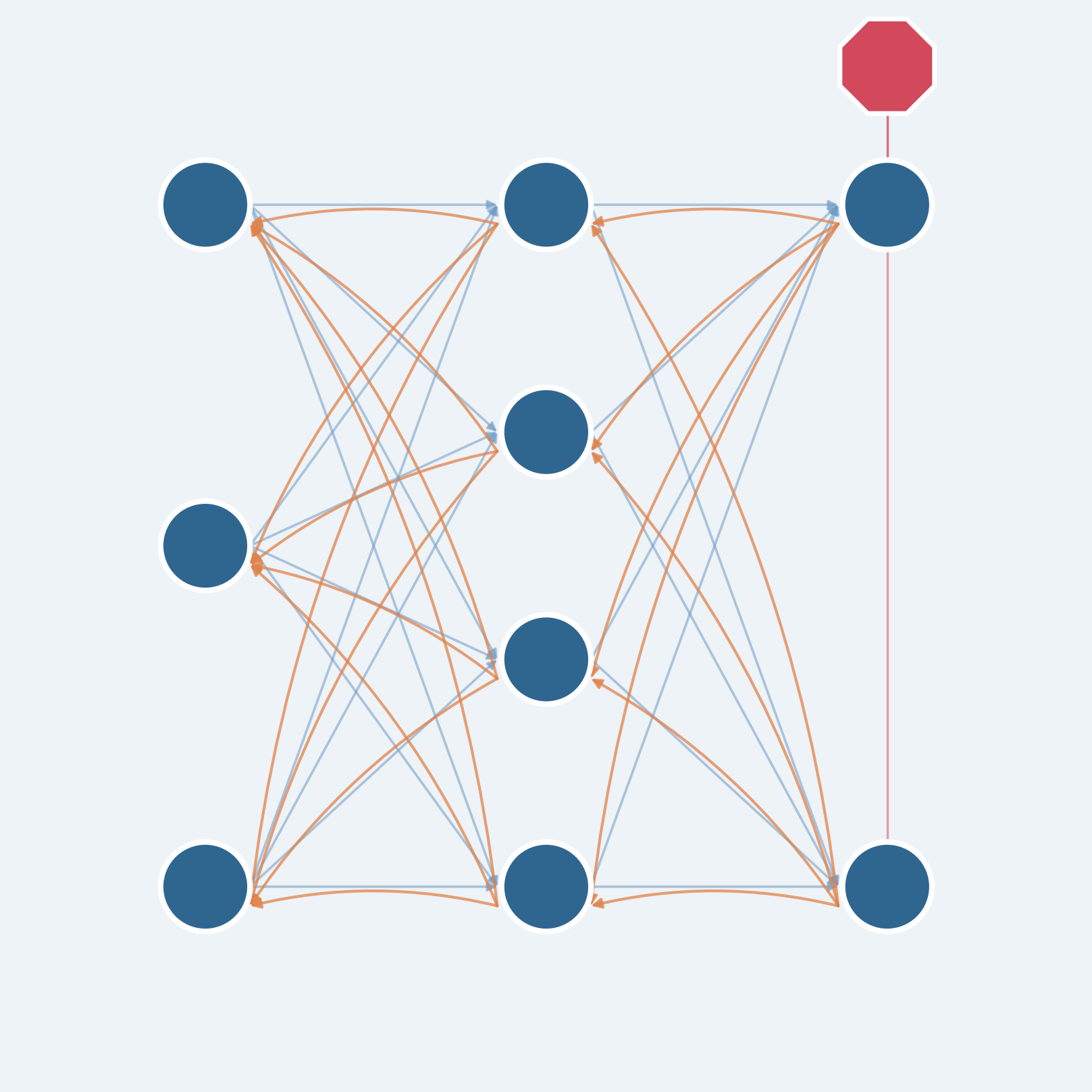
    Training with backpropagation happens in two passes. In the forward pass, an input — a photo, a sentence, a row of sensor data — moves through the network’s layers in order, and the network produces a prediction. That prediction is then compared with the correct answer using a loss function, a numerical measure of how far the prediction was from the desired result.

    Backpropagation doesn’t tell a network what to think. It tells the network how wrong it was, and in which direction each connection should move to be a little less wrong next time.

    A simple analogy helps before the math: imagine a product moving through several stages of a factory line, and the finished item turns out defective. To find the cause, managers work backward from the end of the line, and each stage reports how much it may have contributed to the problem, passing that information to the stage before it. The backward pass in backpropagation does something similar, except it uses calculus to work this out exactly. Starting at the output layer and moving back toward the input, the algorithm applies the chain rule — a technique for tracking how a change at one stage ripples through a chain of dependent calculations. Each layer combines information about how its own calculations affected the next layer with the error information passed back from that layer, producing a gradient: a measure of how sensitive the loss is to a small change in each weight, at its current value.
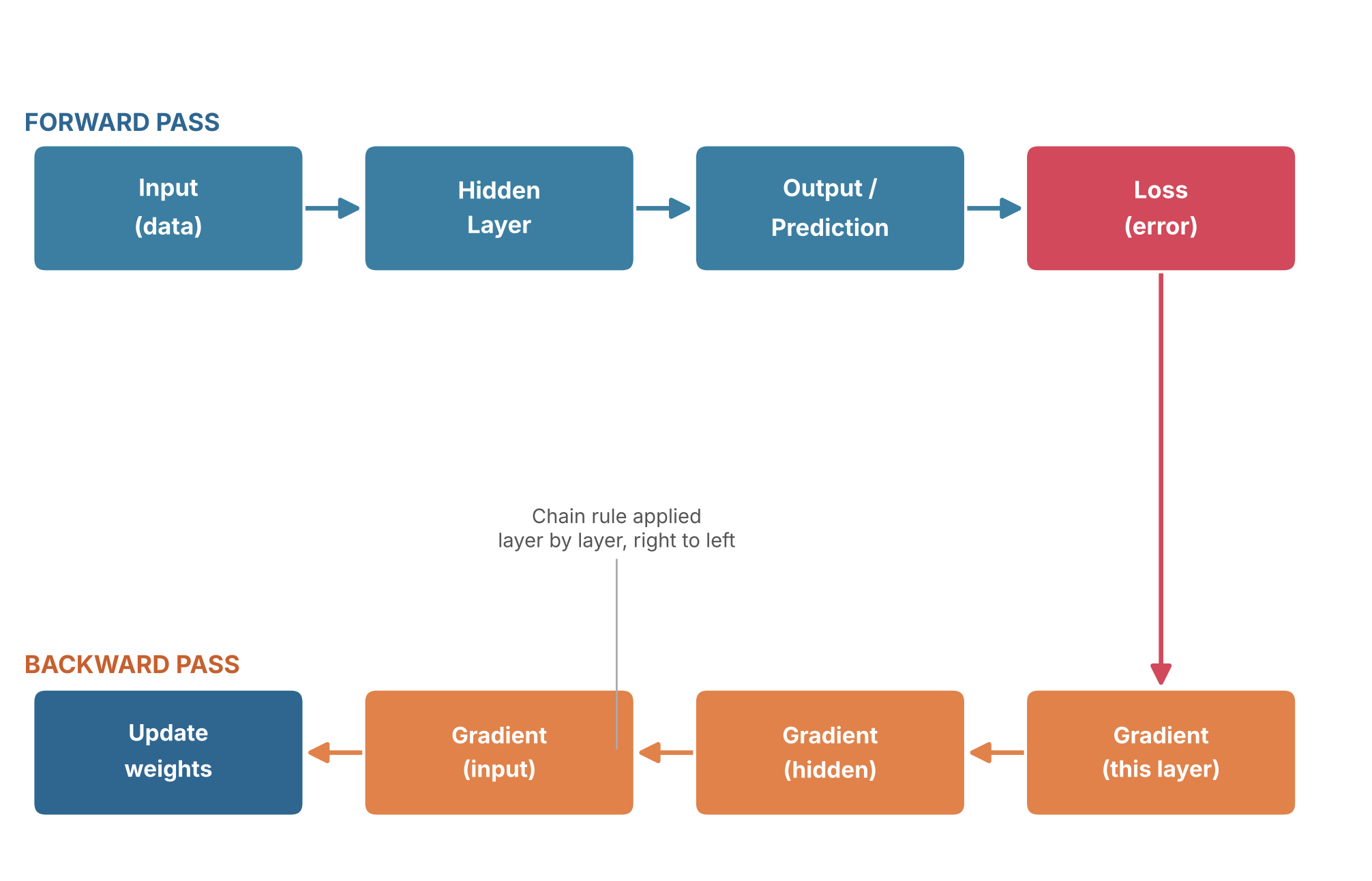
    Diagram of the forward pass and backward pass in backpropagation
    The forward pass produces a prediction and a loss; the backward pass applies the chain rule, layer by layer, to compute gradients and update the weights.

    Once every weight has a gradient, gradient descent uses it to actually update the weights, nudging each one a small amount in the direction that reduces the error. Repeat this forward-pass/backward-pass/update cycle over thousands or millions of examples, and a network that started with randomly initialized connections gradually comes to reflect real structure in the data. The appeal of backpropagation is efficiency: it calculates gradients for every weight in a network in roughly the time needed for a small number of forward and backward passes, rather than requiring a separate experiment for each individual weight.

    Why Backpropagation Is Getting Attention Again

    Backpropagation is not new, and its mathematical core had already been explored before it became famous. Reverse-mode automatic differentiation — the calculus technique at the heart of the algorithm — traces back to Finnish researcher Seppo Linnainmaa’s 1970 master’s thesis, and American researcher Paul Werbos proposed applying a related idea to neural networks in his 1974 doctoral thesis. The method became particularly influential after David Rumelhart, Geoffrey Hinton and Ronald Williams published “Learning representations by back-propagating errors” in Nature in 1986, demonstrating that it could let multilayer networks automatically discover useful internal features rather than relying entirely on engineers to design them by hand.

    For years afterward, backpropagation-trained networks were mostly a research curiosity, limited by available data and computing power. That changed dramatically in 2012, when Alex Krizhevsky and Ilya Sutskever, working with their advisor Geoffrey Hinton, combined backpropagation, the large labeled ImageNet dataset, and graphics-card computing power in a network known as AlexNet. It dramatically outperformed competing approaches in that year’s ImageNet image-recognition competition, a result widely credited with triggering the modern deep learning boom. The large language models behind today’s generative AI chatbots are, at their core, still trained with variations of the same algorithm.
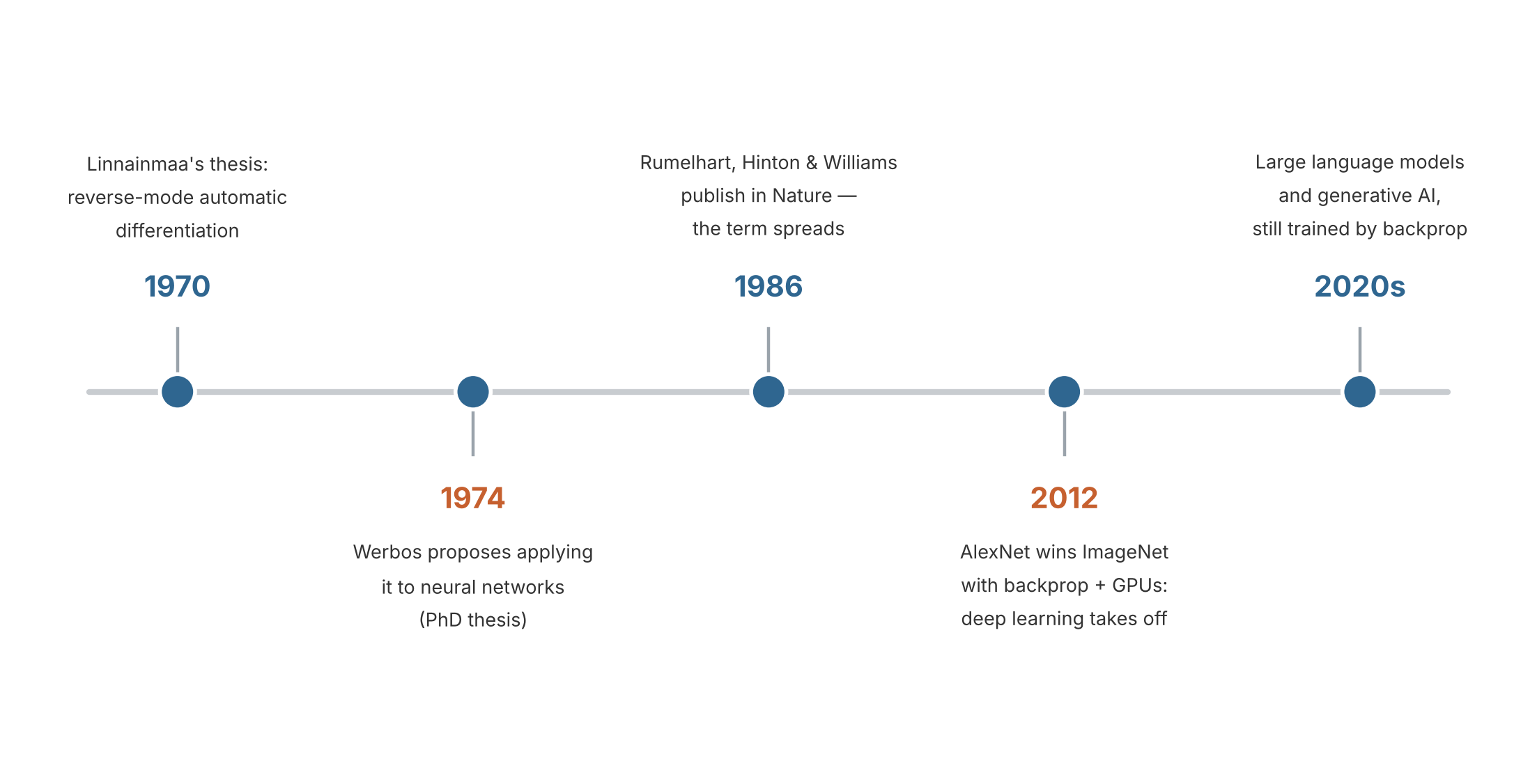
    Timeline of key milestones in the history of backpropagation, from 1970 to the 2020s
    Source: created based on Linnainmaa (1970), Werbos (1974), Rumelhart, Hinton & Williams, Nature (1986), and the AlexNet/ImageNet milestone (2012).

    What has put backpropagation back in the spotlight recently is a different question: does the brain do anything like this? In October 2026, a team at MIT’s McGovern Institute, led by Qianli Liao, Liu Ziyin and Yulu Gan with senior authors Mark Harnett and Tomaso Poggio, published a study in the Proceedings of the National Academy of Sciences proposing a computational model in which randomly connected artificial networks can self-organize their own forward and feedback pathways, using only local learning rules inspired by biological synapses and no global error signal — and still learn nearly as well as backpropagation on the tasks tested. The researchers are careful to frame this as a candidate mechanism and a testable hypothesis, not proof that real brains work this way, but it reopens a question that has nagged neuroscientists for roughly 40 years: backpropagation is clearly powerful enough to drive today’s AI, yet nothing in a biological brain has been shown to do precisely what it requires. (Related article: A Simulated Brain Circuit Builds Its Own AI-Like Learning System, MIT Finds.)

    Where You’ve Already Met Backpropagation

    Even though the math stays behind the scenes, the results of backpropagation-trained networks are part of ordinary life. Many modern face-unlock features and the photo apps that automatically group pictures of the same person run on neural networks trained with backpropagation or related gradient-based methods. Voice assistants that transcribe speech or answer spoken questions generally rely on networks trained the same way. Generative AI chatbots are also built using neural networks trained with backpropagation — during an early stage called pretraining, for example, a language model repeatedly tries to predict the next word in a passage of text, compares its guess with the actual word, and adjusts its weights to reduce the error; later training stages then refine how it follows instructions and responds to feedback.

    It’s worth keeping one distinction in mind: backpropagation mainly happens during training, not every time you use these tools. Once a network has learned, it typically uses its already-adjusted weights to make predictions, without recalculating gradients or updating itself each time — the heavy lifting happened earlier, often across billions of training examples.

    What to Watch Next

    Backpropagation itself is a mature, well-understood technique, so the open questions now sit in its limitations and the alternatives researchers are exploring. One is scientific: whether the MIT team’s self-organizing, locally-plausible learning circuits — or related approaches sometimes called feedback alignment or predictive coding — can be tested directly against real neural tissue, and whether they can match backpropagation’s performance on problems much larger than the ones tested so far. Another is practical: as AI models keep growing, researchers continue to look for training methods that need less computing power than standard backpropagation while keeping its accuracy. The next breakthrough may not come from making backpropagation itself faster, but from discovering how networks can learn without depending on it so completely — which is exactly the question AI researchers and neuroscientists are now asking together.

    Xsciens Editorial Note

    Backpropagation is best understood as a precise bookkeeping trick for calculating how a network should adjust itself, not as a theory of how any brain, artificial or biological, actually “thinks.”

    • A common misconception. “Neural network” learning is often described as though it copies how the brain learns, since artificial “neurons” were loosely inspired by biological ones. In practice, standard backpropagation’s backward calculation relies on precise weight-related information tied to the exact connections used for forward signaling — something no real neuron is known to reproduce or transmit in the same way. (Some alternative methods, such as feedback alignment, relax this requirement, which is itself a sign of how specific the original objection is.) The resemblance between artificial and biological “neurons” is a loose analogy, not a working description of what either one computes.
    • Limits and open questions. A common misreading is that backpropagation always needs a human-provided “correct answer,” like a teacher grading homework. That’s not quite right: it needs a differentiable training objective, which can come from supervised labels, but also from self-supervised tasks (such as predicting the next word in a sentence) or reinforcement-style rewards. What backpropagation does not explain, on its own, is why some network architectures generalize well to new data and others don’t, or how an AI system might decide what is worth learning in the first place — those remain active, only partly answered research questions, not settled ones.
    • What to watch next. The MIT team’s October 2026 study proposes, as a specific and testable hypothesis, that a four-synapse wiring motif linking ascending and descending pathways in the cortex could let real circuits self-organize a similar learning ability. This is a model prediction, not a confirmed feature of brain tissue — whether neuroscientists actually find that wiring pattern, and whether locally-plausible learning rules can match backpropagation’s performance on problems far larger than the ones tested so far, are the two developments worth watching.
    • From the editors — If brain-inspired, locally-learning AI matures the way this MIT team hopes, the assistants people use every day could stop feeling like blank tools that need re-explaining each time and start quietly adapting to how each person actually works. Getting there would likely mean systems that keep adjusting themselves after they leave the lab, learning from ordinary daily use instead of one big retraining cycle. That same ability to keep learning on its own is exactly what to watch: without a way to check what a system has quietly picked up, it could just as easily drift toward learning the wrong lesson from unusual or misleading experience. This is still an early computational demonstration, not a finished product — so if an assistant ever starts to feel like it “gets” you a little faster, it’s worth asking, every so often, what it actually learned to get there.

    References for this note: Quanta Magazine — Artificial Neural Nets Finally Yield Clues to How Brains Learn, arXiv — Brain-Inspired Machine Intelligence: A Survey of Neurobiologically-Plausible Credit Assignment

    Sources: IBM — What Is Backpropagation?, TechTarget — What Is a Backpropagation Algorithm?, Rumelhart, Hinton & Williams, Nature (1986), Seppo Linnainmaa, Computer History Museum — AlexNet Source Code, Xsciens — A Simulated Brain Circuit Builds Its Own AI-Like Learning System, MIT Finds
  • [Science] The Brain’s Alertness System Has More Specific Wiring Than Scientists Thought

    [Science] The Brain’s Alertness System Has More Specific Wiring Than Scientists Thought

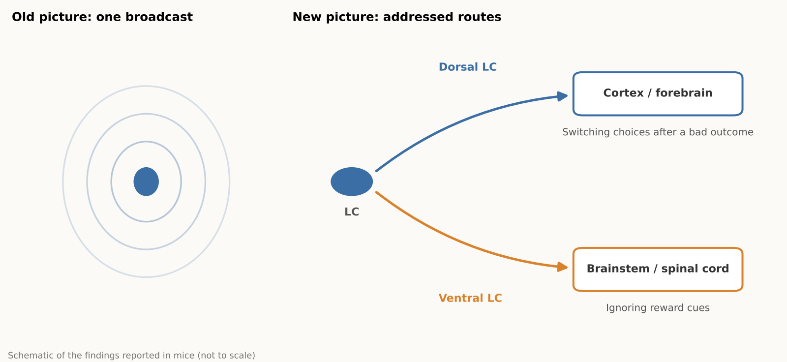
    Norepinephrine, a brain chemical involved in alertness, attention and stress responses, comes largely from a tiny cluster of cells in the brainstem called the locus coeruleus (LC). For decades this system was often pictured as a foghorn, broadcasting much the same signal everywhere. A new Nature study in mice, led by researchers at the Allen Institute, finds a more organized picture, closer to a postal network: different LC neurons tend to send norepinephrine to different destinations and show different activity during behavior. Why it matters: some medications for depression and ADHD influence norepinephrine signaling, and a wiring map is a first step toward understanding how the system could one day be influenced more selectively.

    原論文情報 / Original Paper
    Title: Topographic structure and function of locus coeruleus noradrenaline neurons
    Authors: Zhixiao Su, Polina Kosillo, Kanghoon Jung et al.
    Journal: Nature (2026)
    DOI: 10.1038/s41586-026-11026-0

    What the locus coeruleus does

    The locus coeruleus (LC) is a tiny brainstem nucleus, often nicknamed the “blue spot,” and the main source of norepinephrine, also called noradrenaline, for much of the brain and spinal cord. The chemical helps regulate arousal, attention and mood, and some medications for depression and ADHD act on norepinephrine signaling. Changes in the LC have also been reported early in Alzheimer’s disease, although the timing and significance of that degeneration are still under study.

    The textbook picture often treated the LC as a broadly broadcasting system, rather like a foghorn. Earlier work had already hinted at more structure. A 2020 review in Nature Reviews Neuroscience, for example, concluded that subgroups of LC neurons can encode specific cognitive processes, and a 2023 eLife study found two main electrical and structural cell types in the mouse LC. The new study maps how those differences in wiring, gene expression and activity are organized across the structure.

    Mapping one tiny structure

    Why map a structure this small? Because a nucleus that reaches almost the entire brain is hard to understand from the outside. Unless researchers know which neurons talk to which regions, it is difficult to say whether a drug, a stress response or a behavior is engaging the whole system or only a piece of it. Anatomy comes first, and function can be tested against it.

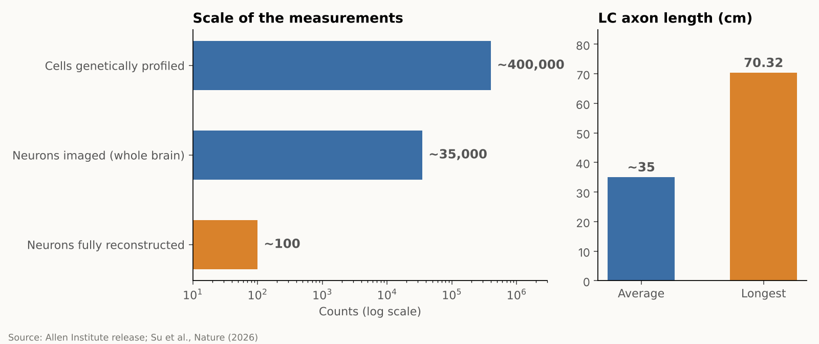
    The team combined several approaches in mice, each answering a different question. Whole-brain imaging showed where LC neurons send their fibers, gene profiling showed which genes they use, and detailed reconstructions traced the branches of individual cells. The researchers then recorded neural activity while mice performed a learning task. In numbers, they imaged almost 35,000 neurons across the whole brain, profiled gene activity in nearly 400,000 cells, and fully reconstructed about 100 individual LC neurons, according to the Allen Institute. These are different measurements from different samples, not 400,000 neurons traced end to end. The research was supported by the NIH BRAIN Initiative.

    Bar charts showing the scale of the measurements and the average and longest LC axon lengths
    Source: Allen Institute news release and Su et al., Nature (2026). Numbers are approximate as reported.

    A map of connections, a gradient of genes

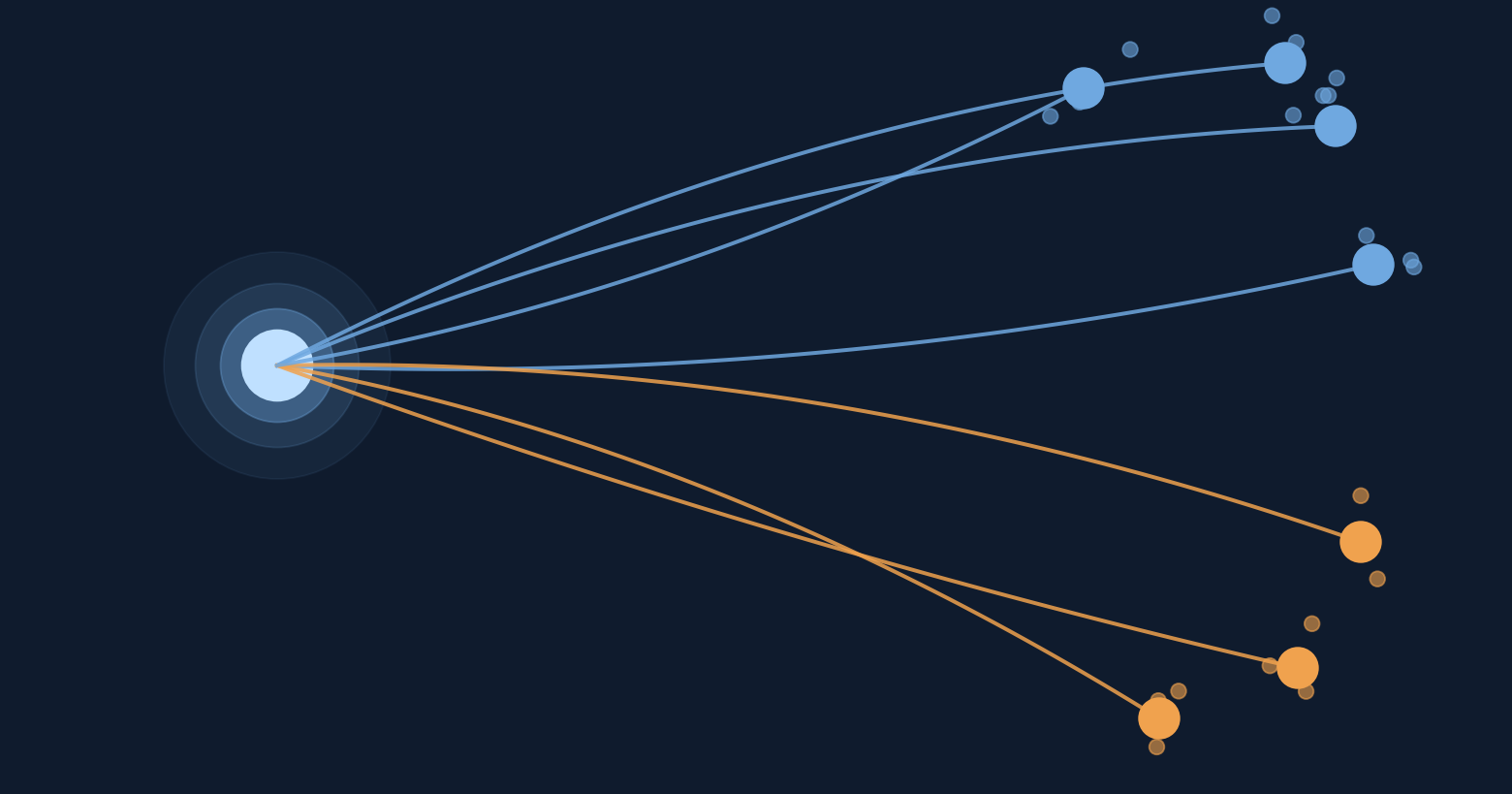
    The reconstructions showed that the LC is organized by position, as a gradient rather than two sealed compartments. Neurons in the upper (dorsal) part mainly send their axons to the cerebral cortex and forebrain, while neurons in the lower (ventral) part mainly project to the brainstem and spinal cord. Gene expression also changes smoothly along that same dorsal-to-ventral axis, matching the projection pattern, according to a summary of the paper’s abstract.

    The striking part is that these patterns line up. A neuron’s position in the LC helps predict where its axon projects and how its gene expression differs from its neighbors, and, in some cases, how it behaves during learning.

    A very long cell, and a selective one

    LC axons, the long fibers that carry signals out of the cell, averaged about 35 centimeters in total length in the mice. The longest one traced measured 70.32 centimeters in total, which the Allen Institute describes as the longest single neuron measured in a mouse. That neuron supplies norepinephrine to a large volume of cortex but does not release it in the cerebellum, brainstem or spinal cord.

    “The findings suggest the brain’s norepinephrine system is far more like a targeted postal network than a foghorn,” said Karel Svoboda of the Allen Institute.

    For readers who follow how the brain learns, this fits a broader theme: learning is not one switch but many coordinated signals. We covered one such idea in our earlier piece on an MIT hypothesis about how the brain might build its own learning circuits.

    Schematic contrasting a single broadcast with two addressed LC routes to cortex and spinal cord
    Schematic based on findings reported in mice. Not to scale.

    Different routes, different jobs

    The activity recordings pointed to different roles. Cortex-projecting dorsal neurons became more active during learning, specifically when mice changed their choice after an unfavorable outcome, and their activity also tracked reward prediction error, the gap between the reward an animal expected and what it actually got, which can guide future choices. Ventral neurons had higher background activity when mice ignored cues indicating that a reward might be available. The authors suggest this may reflect differences in how animals respond to potentially relevant cues.

    A signal that carries that gap is a natural teaching tool. Finding one in a norepinephrine pathway, rather than only in the better-known dopamine system, is why the authors are interested in how the two might work together. They suspect norepinephrine and dopamine may supply complementary learning signals, but that remains a hypothesis. According to The Transmitter, the team plans to study how these chemicals interact next.

    Researchers who were not involved in the work have commented on an earlier preprint version of the study. Nelson Totah of the University of Helsinki called it “bread-and-butter” research for the field that answers basic questions about an evolutionarily ancient structure. Rebecca Jordan of the University of Edinburgh said it suggests the LC could send tailored information to different brain areas, and Robert Froemke of NYU noted that the gene-expression profiling could help researchers record and manipulate specific cell types, according to The Transmitter. The published Nature paper was not evaluated by these experts in that coverage.

    An independent 2025 review adds some context. It summarizes rodent studies in which ventral LC neurons reduced pain through projections to the spinal cord, while activating the LC–prefrontal circuit increased pain and anxiety-like behavior. These results come from separate experiments, but they support the idea that LC pathways can have different effects depending on their destinations.

    Why it matters, and what to keep in mind

    Because norepinephrine-related drugs usually act across the whole brain, a map of separate pathways raises the possibility of more selective ways to influence them that avoid unrelated circuits. The Allen Institute describes this as a possibility for the future, not a demonstrated result. All of the work was done in mice, and nobody has yet shown that human LC neurons are organized the same way. Neither does the study show that any of these pathways can be treated on its own. For now, it is a map, and a good one, rather than a medical advance.

    The practical takeaway for a general reader is modest but real: the brain’s alertness and attention system is more finely organized than the old “one signal for everyone” picture suggested. That kind of basic knowledge tends to pay off slowly, by giving later researchers better targets and better questions.

    Xsciens Editorial Note

    The study suggests that a brain system once treated as a single volume knob is better understood as a network with different destinations. If that view is confirmed in people, treatment for focus and mood, learning support and early detection of brain aging could shift from adjusting the whole brain to targeting only the wires that matter, which may shape the precision of future drugs and diagnostics.

    • What is solid is the map; what is still a hypothesis is the job of each route. Where the axons go, how gene use shifts along the structure, and the recorded activity are direct measurements, so any past interpretation that assumed one broadcast signal now needs revisiting. Reading the dorsal neurons as a “learning signal,” or the ventral ones as tied to responses to reward cues, is an interpretation drawn from how activity lines up with behavior, and the norepinephrine–dopamine partnership remains a hypothesis. These are the claims coming experiments will test.
    • A mouse map is a blueprint; confirming it in people is the next stage. About 100 neurons were reconstructed in full, according to the Allen Institute, and the 35,000 imaged and 400,000 profiled cells are separate measurements. An independent 2025 review also notes that much of the evidence comes from rodents. The flip side is useful: the map tells researchers exactly what to look for in human tissue, which is where the field has to go.
    • Three kinds of study to expect next (our projections). (a) Causal experiments that silence or activate one pathway at a time and see whether behavior changes, a question the study leaves open. (b) Simultaneous recordings of norepinephrine and dopamine to test whether they divide the work of learning, which The Transmitter reports the team plans to study. (c) Studies in human brains, such as post-mortem tissue or imaging, that look for the same dorsal–ventral arrangement.
    • From the editors — the idea of raising or lowering a brain chemical across the board may give way to tuning one wire at a time. This map points to three futures.
      1) Drug design. Today’s norepinephrine-related drugs move the pathway that serves focus and the pathway that drives bodily arousal together. The finding that gene use shifts gradually from dorsal to ventral gives researchers a possible marker for telling routes apart, and an outside expert quoted by The Transmitter noted that gene profiling could help record and manipulate specific cell types. A treatment that nudges the focus-related part without the racing heart and tension is a design goal this map makes more realistic.
      2) Understanding learning. Dorsal neurons responded when mice switched choices and when outcomes missed expectations, which suggests this system may help teach us at the moment we recover from a mistake. If so, the best timing of feedback could become a design question for teaching and rehabilitation. For AI research, the idea of separating a “learn the value” signal from a “change your approach” signal into different wires may be worth borrowing.
      3) Spotting change early. Changes in the LC have been reported early in Alzheimer’s disease. With this map as a ruler, researchers could ask which pathways are damaged first and whether those changes can be caught earlier and in finer detail. This study did not test that.
      These are our projections, and the road from a mouse map to people is still ahead. One way to read the news in the meantime: when you see a headline about a drug that targets a particular brain pathway, check which pathway was tested in humans. That is where a map turns into a treatment, or does not.

    References for this note: McKinney et al., eLife (2023), Hao et al., Int. J. Mol. Sci. (2025), The Transmitter (2026), Poe et al., Nature Reviews Neuroscience (2020)

    Sources: Allen Institute, Su et al., Nature (2026), ScienceDaily

  • [Science] What Is Cognitive Offloading? A Simple Guide to How It Works

    [Science] What Is Cognitive Offloading? A Simple Guide to How It Works

    Every time you set a phone reminder instead of memorizing an appointment, or ask a chatbot to summarize a report instead of reading it yourself, you are doing something psychologists call cognitive offloading. The habit itself is ancient, but a wave of recent research is asking a sharper question: what happens to our own performance in the moments when the tool isn’t there?

    Source Paper
    Title: Cognitive Offloading
    Authors: Evan F. Risko, Sam J. Gilbert
    Journal: Trends in Cognitive Sciences, Vol. 20, No. 9, pp. 676–688 (2016)
    DOI: 10.1016/j.tics.2016.07.002

    What Is Cognitive Offloading?

    Cognitive offloading is the use of physical action or an external tool to reduce the mental effort a task would otherwise require. Psychologists Evan Risko (University of Waterloo) and Sam Gilbert (University College London) published an influential review of the concept in 2016, in Trends in Cognitive Sciences, which remains the field’s clearest framework for the idea. Rather than solving a problem entirely “in your head,” you shift part of the work onto your body or your environment.

    It’s a far older behavior than smartphones. Tilting your head to read an upside-down sign, counting on your fingers, or jotting a phone number on a scrap of paper are all forms of cognitive offloading. What has changed is the scale and power of the tools available — calculators, GPS, search engines, and now generative AI assistants can take over not just small chores but entire chains of reasoning.

    The idea is closely related to the “extended mind” thesis, proposed by philosophers Andy Clark and David Chalmers in 1998. They argued that a notebook carried everywhere and consulted automatically can function, for practical purposes, as part of a person’s memory system — their famous example involved a man named Otto who used a notebook the way most people use their own recall. Risko and Gilbert’s later work gave this philosophical idea an empirical, testable framework: when exactly do people choose to offload, and what happens to their underlying skills when they do it repeatedly?

    How It Works: A Decision Loop, Not a Switch

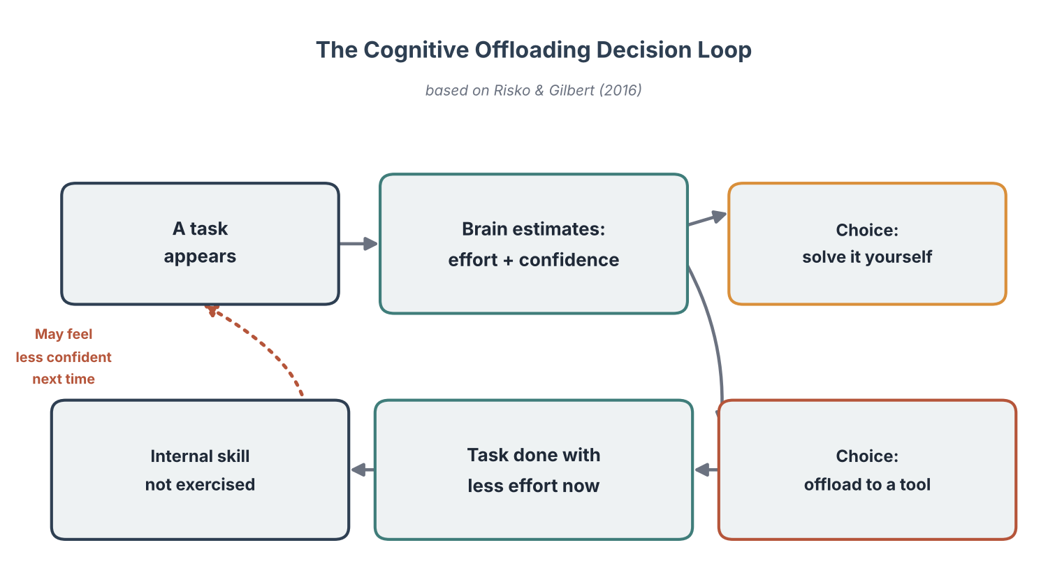
    Imagine being asked to remember five errands on the way home. You could hold them all in mind, or you could write them down. Risko and Gilbert’s framework treats this as a running cost-benefit calculation rather than a one-time choice: the decision depends on how demanding the task feels, how confident you are in your own memory, and how much effort writing the list would save.

    The important detail is that this calculation runs on perceived demand and confidence, not necessarily on actual ability — and people’s self-assessments can be mistaken, so they sometimes reach for an external aid even when they could have managed without it. Whether repeated reliance on a tool gradually changes a person’s confidence or unaided performance over the longer term is a related but separate question, one that is harder to answer than the moment-to-moment decision itself. Offloading isn’t inherently a problem — compensating for a busy week with a written list is a perfectly sensible trade — but it is a trade worth noticing rather than making automatically.

    Diagram of the cognitive offloading decision loop
    Source: created based on Risko & Gilbert (2016), Trends in Cognitive Sciences

    Why It’s Back in the Spotlight Now

    Cognitive offloading has been studied for decades, but generative AI has pushed it from a niche academic topic into a mainstream concern. A widely discussed 2011 study in Science by Betsy Sparrow, Jenny Liu, and Daniel Wegner found that once people believed information would remain accessible online, they remembered where to find it far better than the information itself — a pattern nicknamed the “Google effect.” That study is a useful historical analogy rather than direct proof of what AI chatbots do to the brain: a search engine mainly stores and retrieves information, while an AI assistant can also perform reasoning-like tasks, draft text, and summarize on a person’s behalf, which is a different kind of delegation that researchers are only beginning to study on its own terms.

    The question researchers are now asking is not whether people offload thinking to AI, but how much of their own judgment they lose when the AI isn’t there.

    A 2025 study published in Societies by Michael Gerlich surveyed 666 participants and reported a statistically significant negative correlation between frequent AI tool use and scores on critical-thinking tasks, with younger, more AI-reliant participants scoring lowest. Because the study measured AI use and critical-thinking ability at a single point in time, it cannot determine whether frequent AI use weakens critical thinking, whether people with different existing abilities simply use AI differently, or whether some other factor explains both — but it adds to a growing body of research raising the same question from different angles.

    Everyday Examples You Already Use

    Cognitive offloading shows up in ordinary moments far more often than most people realize. A few familiar examples: writing a shopping list instead of memorizing items, letting a smartphone calendar track every appointment, typing a destination into GPS navigation instead of memorizing the route, and asking a search engine a factual question instead of trying to recall it. In most of these cases the trade is a fair one — a shopping list, for instance, tends to help you finish the errand without costing you much else. The more interesting question researchers are asking is which kinds of offloading are closer to that fair trade, and which carry a greater cost to independent performance later on.

    A 2020 study in Scientific Reports, led by Louisa Dahmani and Véronique Bohbot at McGill University, examined this trade-off directly with GPS. In a sample of 50 regular drivers, heavier GPS use was associated with weaker spatial memory on unaided navigation tasks. A smaller follow-up of 13 of the same participants about three years later found that people whose GPS use had grown the most over that period also showed the steepest declines in spatial memory — a dose-response pattern the authors say is more consistent with GPS use contributing to the decline than the reverse, though they are careful to flag their small sample size and note that the direction of cause and effect is not fully settled.

    Not every example involves memory. Risko and Gilbert’s framework also covers offloading onto the body itself — pointing at an object instead of holding its location in mind, or using a finger to keep a place while reading a long list — and “intention offloading,” where a sticky note or phone alert carries the job of remembering to act later, rather than the job of remembering the information itself. A related but distinct idea is automation, where a system completes part of a task with no decision from the user at all. A GPS app that both tracks your route and reroutes you around traffic without being asked is doing more than simple offloading; it raises its own, separate questions about what happens to a skill when a person is never required to exercise it in the first place.

    What Recent Research Shows

    The clearest recent demonstration comes from a UC Berkeley–led study published in 2026. Researchers ran three randomized trials with 1,222 participants who solved math and reading problems either with or without ChatGPT. Everyone then had to solve a final set of problems entirely unaided. Participants who had previously relied on the AI scored markedly lower on those unaided problems than participants who had worked alone from the start — in one trial, 57% versus 73% correct. Related article: AI Assistance Reduces Persistence and Hurts Independent Performance looks at this study’s full results and the researchers’ own caveats about how far the findings generalize.

    None of this means offloading is simply bad. In a self-reported, non-randomized part of the same study, participants who said they had copied AI-generated answers directly scored the lowest on the unaided follow-up problems (65% correct on average), while participants who said they had used the AI only for hints scored close to those who never used AI at all. Because that comparison is based on what participants reported about their own behavior rather than a randomized condition, it should be read as a suggestive pattern rather than a proven result. Still, it points toward a recurring theme across this research: the question is less whether to offload and more how — leaning on a tool for a finished answer looks different, in its effects, from using it as a scaffold while you still do the thinking yourself.

    Sources: Risko & Gilbert, “Cognitive Offloading,” Trends in Cognitive Sciences (2016), Gerlich, “AI Tools in Society,” Societies (2025), Columbia News on Sparrow et al., Science (2011), American Psychological Association Monitor (2026)

    See also: Why Letting AI Help Can Erode Your Grit and AI Summaries Can Distort Human Memory.

  • A Simulated Brain Circuit Builds Its Own AI-Like Learning System, MIT Finds
    A Simulated Brain Circuit Builds Its Own AI-Like Learning System, MIT Finds
    For decades, neuroscientists have faced a nagging puzzle. Modern AI owes its power to backpropagation, an algorithm that adjusts a network’s connections by propagating error information backward through precisely coordinated connections — a computation with no clearly established equivalent in biological circuitry. Yet some studies have found that activity patterns in trained artificial networks resemble those recorded in the brain’s own visual system, which only sharpens the question of how biological neurons, using only local signals and changing synapses, might approximate something similar. A new study from MIT’s McGovern Institute for Brain Research, published October 9 in the Proceedings of the National Academy of Sciences (PNAS), offers a candidate answer: when simple neuron-like units are wired randomly and left to follow local, biologically plausible plasticity rules, they can spontaneously self-organize into circuits that learn nearly as well as backpropagation does on the tasks tested — without anyone telling them how.

    原論文情報 / Original Paper
    Title: How Biological Synapses Self-Assemble Gradient Learning
    Authors: Qianli Liao, Liu Ziyin, Yulu Gan, et al.
    Journal: Proceedings of the National Academy of Sciences (PNAS)
    DOI: 10.1073/pnas.2606245123

    A 40-Year Mismatch Between Brains and AI

    Modern deep learning owes almost all of its success to backpropagation: a network makes a prediction, compares it with the correct answer, and then propagates that error backward, layer by layer, nudging every connection weight in the direction that would have reduced the mistake. It works brilliantly, but standard implementations depend on precise mathematical relationships between forward and backward connections, plus coordinated transmission of error information through the network. There is no established evidence that cortical circuits implement anything equivalent in its conventional form.

    The puzzle deepened as deep learning matured. Comparisons between the internal activity of backpropagation-trained networks and recordings from biological visual systems have repeatedly found intriguing similarities — not identical, but notably closer than chance would predict. Such similarities do not prove the brain runs backpropagation, but they raise an obvious question: if it does not, what local process could still produce computations that end up looking so much like it?

    Local Rules, No Blueprint

    The MIT team, led by Qianli Liao, Liu Ziyin and Yulu Gan with senior authors Mark Harnett and Tomaso Poggio (and co-author Brian Cheung), approached the question differently from most prior attempts to make backpropagation “more biological.” Instead of designing a circuit by hand and checking whether it could plausibly exist in a brain, they started from almost nothing: layers of neuron-like units connected at random, with no predetermined wiring diagram at all.

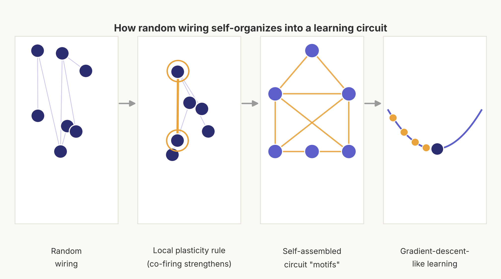
    The system runs on local synaptic plasticity rules, including heterosynaptic mechanisms that let a connection change in response to activity beyond that single synapse — a more specific relative of the familiar Hebbian idea that “cells that fire together wire together.” Critically, these rules only need information available locally, at or near a given synapse (the junction where one neuron signals to the next). No unit needs to know what is happening elsewhere in the network, and no single global error signal is broadcast to the whole system.

    Diagram showing random wiring self-organizing into a learning circuit
    Source: created based on findings from MIT McGovern Institute / PNAS (2026).

    Circuits That Wire Themselves

    Left to run under those local rules, the randomly connected units did not stay random for long. Over repeated rounds of activity, the network developed small, recurring wiring patterns the researchers call “self-assembling motifs.” The model’s analysis points to a specific four-synapse motif linking the brain’s ascending, bottom-up pathways with its descending, top-down pathways — a concrete structural prediction that experimentalists can now go looking for in real neural tissue, not a pattern already confirmed to exist there.

    What these motifs do next is the more remarkable part: networks of them self-organize further, so that their combined dynamics approximate a generalized form of stochastic gradient descent — the family of optimization methods that includes backpropagation — without a hand-designed, globally coordinated algorithm directing them. In other words, before the network learned anything from its training data, it first had to build the machinery that would let it learn.

    Starting from random connectivity, local synaptic plasticity rules were enough for the network to assemble circuit motifs whose combined dynamics approximate gradient-descent-style learning.

    Holding Its Own Against Backpropagation

    A biologically plausible circuit is only interesting if it actually works, so the researchers tested their self-organized networks on image pattern recognition and classification tasks. According to MIT, the self-assembled circuits achieved performance comparable to conventional networks trained directly with backpropagation on the tasks tested. The MIT News announcement does not report specific accuracy figures or benchmark datasets — those details sit in the full PNAS paper — so the result is best read as evidence that self-organization carried no obvious cost on this particular test, not as a claim that it matches backpropagation across deep learning in general.

    That matters because it suggests biological plausibility and computational competence are not necessarily in tension. For years, biologically inspired alternatives to backpropagation have often underperformed it on real tasks. A circuit that assembles itself from random wiring and still competes with backpropagation on the tasks it was tried on narrows that gap, at least in this one demonstration.

    A Prediction Neuroscientists Can Go Test

    The researchers are careful to note that a self-organizing network learning like this does not prove the human brain learns this way — it is a candidate mechanism, not a confirmed one. But unlike many theoretical proposals in computational neuroscience, this one comes with a concrete, falsifiable prediction: if the brain really does something like this, cortical circuits should contain that specific four-synapse motif connecting ascending and descending pathways. Experimental neuroscientists can now search actual brain tissue for exactly that wiring signature, which is what turns an elegant simulation into a testable scientific hypothesis.

    Why It Matters Beyond the Lab

    If the prediction holds up, the payoff would extend in both directions. For neuroscience, a confirmed biologically plausible learning circuit would help explain how the brain adapts throughout life, and could sharpen our understanding of what goes wrong in conditions involving altered learning or memory, as well as inform the design of future brain-computer interfaces that need to interact with the brain’s own learning rules rather than fight against them.

    For AI, the lesson cuts the other way: today’s networks are built top-down, with every connection and training rule specified by engineers before a single example is seen. A system that can bootstrap its own learning machinery from random wiring and local plasticity rules hints at an alternative path — architectures that configure themselves as they encounter data, rather than ones that are handed a finished blueprint. Liao, Ziyin, Gan and their colleagues frame their result as an early step in that direction, not a finished product, but their work gives both neuroscientists and AI researchers a concrete new question to test: not just how networks learn once they are built, but how learning itself can come preinstalled.

    For more on where artificial and biological intelligence still diverge, see our earlier coverage of how MIT neuroscientists found the brain re-files information the instant you make a decision, a separate finding that is likewise reshaping how AI researchers think about adaptive, flexible learning.

    Sources: MIT News, Proceedings of the National Academy of Sciences (PNAS), arXiv preprint

  • [Science] MIND Diet Linked to Years of Slower Brain Aging, 12-Year Study Finds

    [Science] MIND Diet Linked to Years of Slower Brain Aging, 12-Year Study Finds

    A diet designed around brain health may actually be protecting it. A 12-year study of nearly 1,650 American adults found that people who more closely followed the MIND diet — a hybrid of the Mediterranean and DASH diets — showed significantly slower shrinkage in brain regions tied to memory and thinking. Researchers calculate the effect as equivalent to roughly 2.5 years of delayed brain aging. The findings, published in the Journal of Neurology, Neurosurgery & Psychiatry, offer some of the strongest long-term evidence yet that everyday food choices in midlife may shape how the brain ages decades later.

    Inside the MIND Diet Study

    The MIND diet (Mediterranean-DASH Intervention for Neurodegenerative Delay) scores people from 0 to 15 based on how often they eat foods linked to brain health, such as leafy greens, berries, nuts, whole grains, fish, and olive oil, while subtracting points for red meat, fried food, sweets, cheese, and butter. For the new study, researchers from Zhejiang University, Harvard T.H. Chan School of Public Health, and the long-running Framingham Heart Study followed 1,647 adults from the Framingham Offspring Cohort, who were around 60 years old on average when the study began.

    Starting in the early 1990s, participants filled out detailed food-frequency questionnaires to calculate their MIND diet scores, with an average score of just under 7 out of 15 — meaning most people in the study were eating a moderately, not perfectly, brain-healthy diet. From 1999 onward, each participant underwent brain MRI scans roughly every few years — three scans on average — allowing researchers to track real changes in brain volume over a median follow-up of 12.3 years, rather than relying on a single snapshot. This repeated-measurement design is part of what makes the study notable: most diet-and-brain research relies on a single MRI scan compared against a single diet survey, which can only show a correlation at one point in time. Tracking the same people’s brains over more than a decade let the researchers instead measure the pace of change itself.

    Years of “Younger” Brain Structure

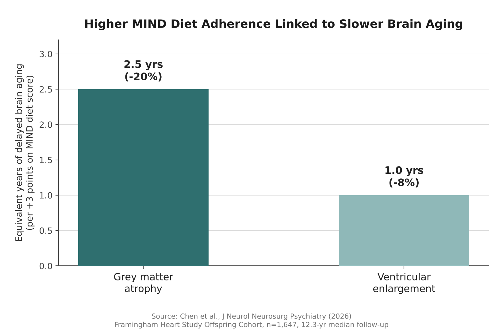
    The results were striking for an observational study. For every 3-point increase in MIND diet score, participants’ grey matter — the brain tissue that handles memory, decision-making, and sensory processing — shrank 0.279 cubic centimeters less per year, about 20% less age-related loss than would otherwise be expected. Researchers translated that gap into real-world terms: roughly 2.5 fewer years of brain aging.

    A 3-point higher MIND diet score was linked to about 2.5 fewer years of brain aging in grey matter volume — one of the clearest signals yet connecting everyday diet to long-term brain structure.

    Higher MIND scores were also linked to slower enlargement of the brain’s ventricles, fluid-filled spaces that expand as surrounding tissue is lost with age. That slower expansion — about 8% less than expected — translated to roughly one additional year of more youthful brain structure. Because all participants’ brains shrank somewhat over the study period, as is normal with aging, the real finding is about the pace of that decline, not whether it happened at all.

    Bar chart showing MIND diet score linked to years of delayed brain aging
    Source: Based on data from Chen et al., J Neurol Neurosurg Psychiatry (2026)

    Foods That Helped, and Surprised

    Not every food in the MIND diet scoring system behaved as expected once researchers looked food by food. Berries were linked to slower enlargement of the ventricles, and poultry was linked to both slower ventricular enlargement and slower grey matter loss — consistent with berries’ high antioxidant content and poultry’s role as a lean protein source, both of which may reduce oxidative stress that damages neurons over time.

    On the other end, sweets and fried fast food were linked to faster ventricular expansion and greater shrinkage of the hippocampus, the brain region most closely tied to memory formation. The researchers pointed to inflammation, unhealthy fats, and advanced glycation end-products — compounds that form when sugar reacts with proteins or fats in food, and which have been separately linked to tissue aging throughout the body — as possible mechanisms.

    Two results, however, ran against expectations. Whole grains, which earn positive points in the standard MIND scoring system, were associated with several unfavorable brain changes in this particular cohort, while cheese, which typically loses points, was linked to favorable ones. The study’s authors flagged both findings as unexpected and said they would need further research to explain, rather than drawing firm conclusions from them. One possibility researchers did not rule out is that people’s reasons for eating more or less of a given food — a doctor’s recommendation to cut dairy after a cardiovascular diagnosis, for instance — could confound a simple food-by-food analysis in ways a single study cannot fully untangle.

    Who Benefited Most

    The association between MIND diet adherence and slower brain aging was not uniform across the group. It was strongest among older participants, who face a faster baseline pace of brain aging and may have more to gain from protective factors. It was also stronger among people who were physically active and those who were not living with obesity, hinting that diet may work alongside — rather than instead of — other healthy habits.

    Participants who ate closer to the MIND pattern also tended to share other characteristics: they were more likely to be women and college-educated, and less likely to smoke or to have type 2 diabetes, high blood pressure, or cardiovascular disease. The researchers statistically accounted for these differences in their analysis, but they illustrate how diet quality tends to travel alongside a broader set of health behaviors.

    What This Study Can’t Prove

    Because this is an observational study, it cannot prove that the MIND diet alone causes slower brain aging. People who eat more healthily in general also tend to differ from others in exercise habits, education, and underlying health conditions, and some of the measured benefit may reflect those overlapping factors rather than diet in isolation. The food data also relied on questionnaires completed in the 1990s, which depend on participants’ memory and are vulnerable to reporting error, and the study could not fully rule out mild cognitive impairment at participants’ first brain scan or track how their diets may have changed over the following two decades.

    The study population was also overwhelmingly White, drawn from a single long-running cohort in Massachusetts, so it remains unclear how well the findings would generalize to more diverse populations. Still, the researchers conclude that their results “reinforce the potential of the MIND diet as a brain-healthy dietary pattern.” Diet is only one piece of the brain-aging puzzle — other recent work, including a study on how the Alzheimer’s risk gene APOE4 scars brain blood vessels, points to genetic and vascular factors working alongside everyday habits. Taken together, the evidence suggests that what people eat in midlife is worth taking seriously as a long-term investment in cognitive health, even before a formal clinical trial settles the question of cause and effect.

    Sources: BMJ Group, Journal of Neurology, Neurosurgery & Psychiatry (PMC)

  • AI Vision Misses an Illusion the Brain Shows — A Gap Worth Testing

    AI Vision Misses an Illusion the Brain Shows — A Gap Worth Testing

    A new study finds that after watching something move, people briefly misjudge exactly where a now-still object is sitting — and a matching shift shows up in signals recorded from monkeys’ brains. The seven AI vision models the researchers tested, built to mimic human sight, did not reproduce that shift at all. Published October 2, 2026 in the journal Current Biology, the work from York University in Toronto does not prove that this gap causes real-world AI vision systems to fail. What it does offer is a precise, repeatable benchmark that engineers building self-driving cars, robots, and other systems that must track a changing scene could use to check whether their software handles recent visual history the way biological vision does.

    Static-image benchmarks, how often a system correctly names or locates an object in a single photo, have long been a mainstay of computer-vision research. But the study’s authors point out that real cameras rarely see the world as a single frozen frame; they see a stream of images shaped by whatever happened a moment earlier. Notably, even a video-based network in the test, built specifically to use multiple frames over time, did not reproduce the shift seen in biological vision. That suggests the gap is not simply about feeding AI more frames, but about a specific kind of recalibration that today’s architectures do not learn on their own.

    A Trick of Motion Fools the Brain

    The effect the researchers studied is related to the classic motion aftereffect, often called the waterfall illusion because it was first described by people who stared at flowing water. After watching motion in one direction — stripes sliding left, for example — a stationary object shown afterward can appear displaced in the opposite direction, even though its pixels have not moved at all. The York University team used this positional bias as a precise, measurable probe into how the brain represents where an object is, rather than treating it as a mere curiosity.

    Testing Monkeys, Humans, and Machines

    The research team, led by senior author Kohitij Kar and first author Elizaveta Yakubovskaya, ran related versions of the same basic test on three very different visual systems. Seventy-nine human volunteers, 35 in a no-motion baseline group and 22 in each of two adaptation-direction groups, briefly saw an object flash on screen for a tenth of a second and clicked where they thought it was. Two rhesus macaques viewed similar sequences while chronically implanted electrode arrays recorded signals from more than 150 neurons in the inferior temporal (IT) cortex, a region of the brain’s object-recognition pathway whose activity also happens to carry information about where an object sits. The same images were then fed into seven widely used computer-vision models, ranging from classic image classifiers such as AlexNet and VGG-16 to a vision transformer and a video-based action-recognition network called SlowFast.

    AI Could Find Objects, Not the Illusion

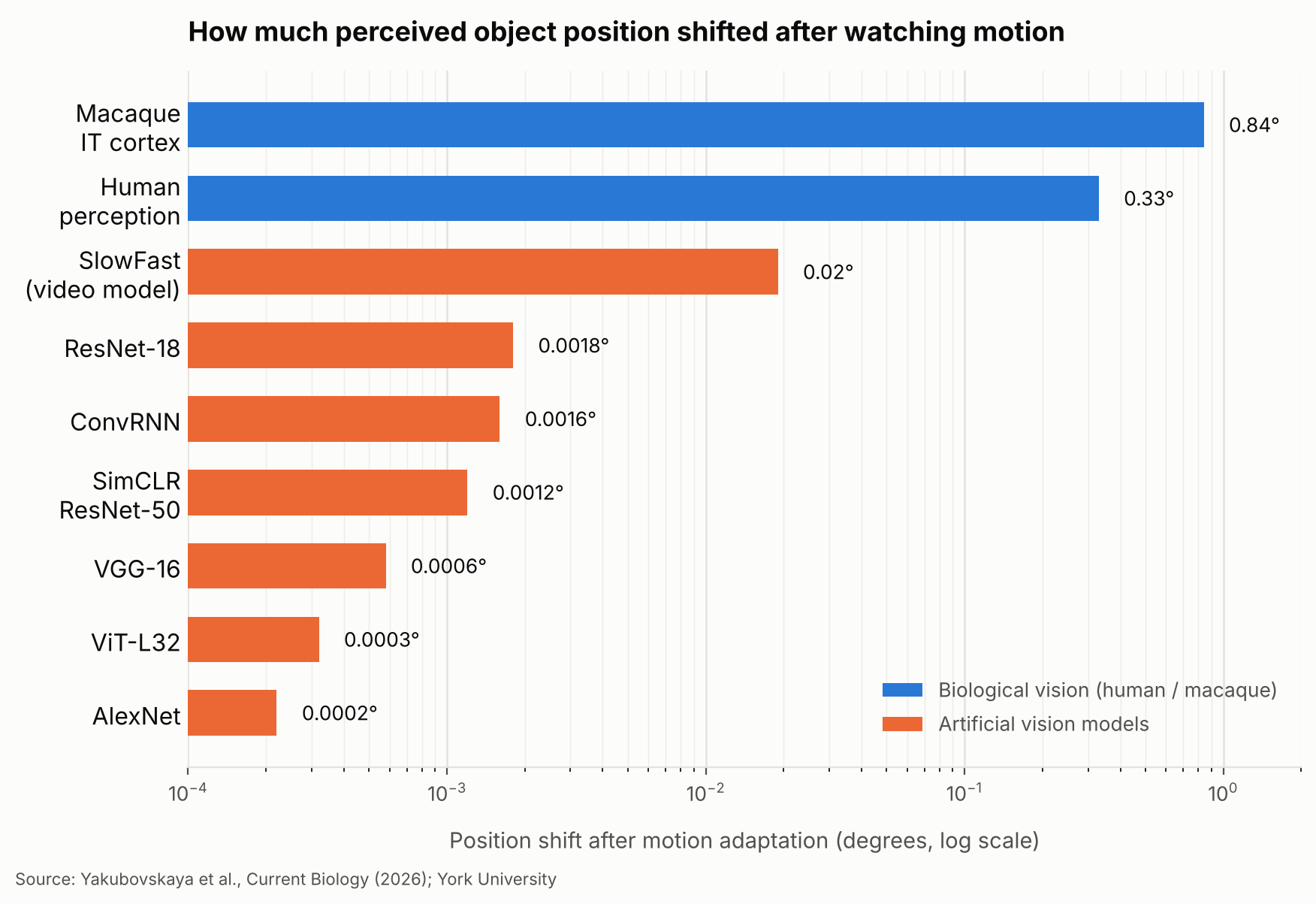
    On ordinary, non-adapted images, both the brain recordings and the AI models could track an object’s true horizontal position: linear readouts from several of the networks correlated with true position about as strongly as readouts from the monkeys’ own IT neurons did, in the range of roughly 0.59 to 0.84. So the models were not simply bad at locating objects. The difference showed up only after adaptation. Human volunteers reported an object’s position shifted by an average of 0.33 degrees of visual angle, opposite the direction of the preceding motion. The position decoded from the monkeys’ IT population activity shifted even further, by 0.84 degrees in the same direction, though that neural shift is a decoded signal rather than a direct report of what the animal consciously saw. Every AI model tested, including SlowFast, shifted by two-hundredths of a degree or less, in practice no meaningful shift at all.

    Human observers reported their perception shifting by 0.33 degrees after watching motion, and the position decoded from monkeys’ IT neurons shifted by 0.84 degrees — while every AI vision model tested shifted by two-hundredths of a degree or less.
    Bar chart comparing object-position shift after motion adaptation across human perception, macaque IT cortex, and seven AI vision models, on a log scale in degrees.
    Source: created based on data from Yakubovskaya et al., Current Biology (2026).

    Copying the Brain’s Adaptation Trick

    The gap did not mean the AI models were incapable of representing position-shifting information at all. When the researchers applied a mathematical transformation, derived from how IT neurons’ responses changed during adaptation, directly onto the models’ internal feature representations, the models began producing human-like position shifts. That shows the models’ feature spaces can support the effect once an appropriate transformation is imposed from outside. It does not show that any of the networks learned to generate that adaptation on their own during ordinary training, which is the gap the authors highlight. This is not the first time researchers have argued that borrowing the brain’s own computations, not just its final answers, could inform AI design; a separate study on how the brain re-files decision options made a related case for mining neural codes rather than just outputs.

    A Gap Worth Testing, Not Yet a Flaw

    The researchers are careful to frame this as a demonstrated difference in representations, not proof that AI vision performs worse in practice. Still, they argue it is a difference worth testing. Self-driving cars, delivery robots, and surveillance systems all judge where things are from a stream of frames in which recent motion could, in principle, matter. Consider a robot tracking a cyclist crossing its camera’s field of view: researchers could test whether the robot’s position estimate shifts the way a human’s perception would after similar recent motion, even though the current image alone has not changed. The York study does not show that reproducing this illusion would make such a system safer or more accurate; it offers a concrete, repeatable way to ask the question. Kar has argued more broadly that judging AI only by whether it reaches the right final answer can miss gaps like this one, and that matching the computations behind human perception, not only matching outputs, may help researchers evaluate whether an AI vision system handles a changing scene the way people expect.

    What the Study Doesn’t Yet Show

    The authors are explicit about the limits of their work. The experiments involved only two macaques and one specific illusion, so it is unclear how far the pattern generalizes to other forms of perceptual adaptation, to more animals, or to other stages of the visual pathway beyond IT cortex. The AI models tested were standard research networks for image and video recognition, not the newer multimodal AI assistants that combine vision with language, which the study did not evaluate. The researchers also do not claim that the perceptual bias they measured is actually beneficial for tracking objects in everyday life, only that it reflects a real computation in primate vision that current AI architectures do not reproduce on their own. They suggest the motion aftereffect could now serve as a simple, repeatable benchmark that other labs and companies use to test how their own vision models handle a changing visual world, and Kar’s group is already pursuing that direction, building what it calls more primate-like artificial neural networks for tasks such as continuous object tracking.

    Sources: Yakubovskaya et al., “The macaque IT cortex but not current artificial vision networks encode object position in perceptually aligned coordinates,” Current Biology (2026), Yakubovskaya et al., arXiv preprint (2026), York University press release coverage, via Neuroscience News

  • [Science] Stimulating the Vagus Nerve After Practice Helps Mice Learn Better Days Later — Tohoku University Finds a Link to Rhythmic Changes in Brain Blood Volume

    [Science] Stimulating the Vagus Nerve After Practice Helps Mice Learn Better Days Later — Tohoku University Finds a Link to Rhythmic Changes in Brain Blood Volume

    • Applying electrical stimulation to the “vagus nerve” on the left side of the neck right after practicing something new may make mice better at it days later, a new study suggests.
    • If a similar effect holds in humans, it could eventually point toward new ways to support motor learning or rehabilitation.
    • Tohoku University researchers stimulated the vagus nerve while tracking local blood-volume changes in the brain, and found that repeated stimulation produced rhythmic fluctuations in local blood volume.
    • Mice with larger blood-volume oscillations after stimulation tended to score higher on a learning test five days later. This is a mouse study, though, and it’s not yet known whether the same effect would occur in humans.

    The Vagus Nerve — a Key Line Between Brain and Body

    The vagus nerve is a major nerve that runs from the brainstem through the neck, chest, and abdomen, carrying signals between the brain and organs such as the heart, lungs, and digestive tract. It’s one of the main nerves connecting the brain and internal organs in both directions: it carries signals about the body’s internal state — such as heart rate, breathing, and digestion — up to the brain, while also carrying signals from the brain back down to those organs.

    A treatment that stimulates this nerve from the outside with electrical pulses is called “vagus nerve stimulation,” or VNS. Using an implanted device, it’s an established therapy for epilepsy that doesn’t respond well enough to medication. In recent years, researchers have also been studying a non-invasive approach that stimulates part of the ear through the skin — though that’s a different method from the one used in this mouse study. Exactly what VNS changes inside the brain has remained unclear.

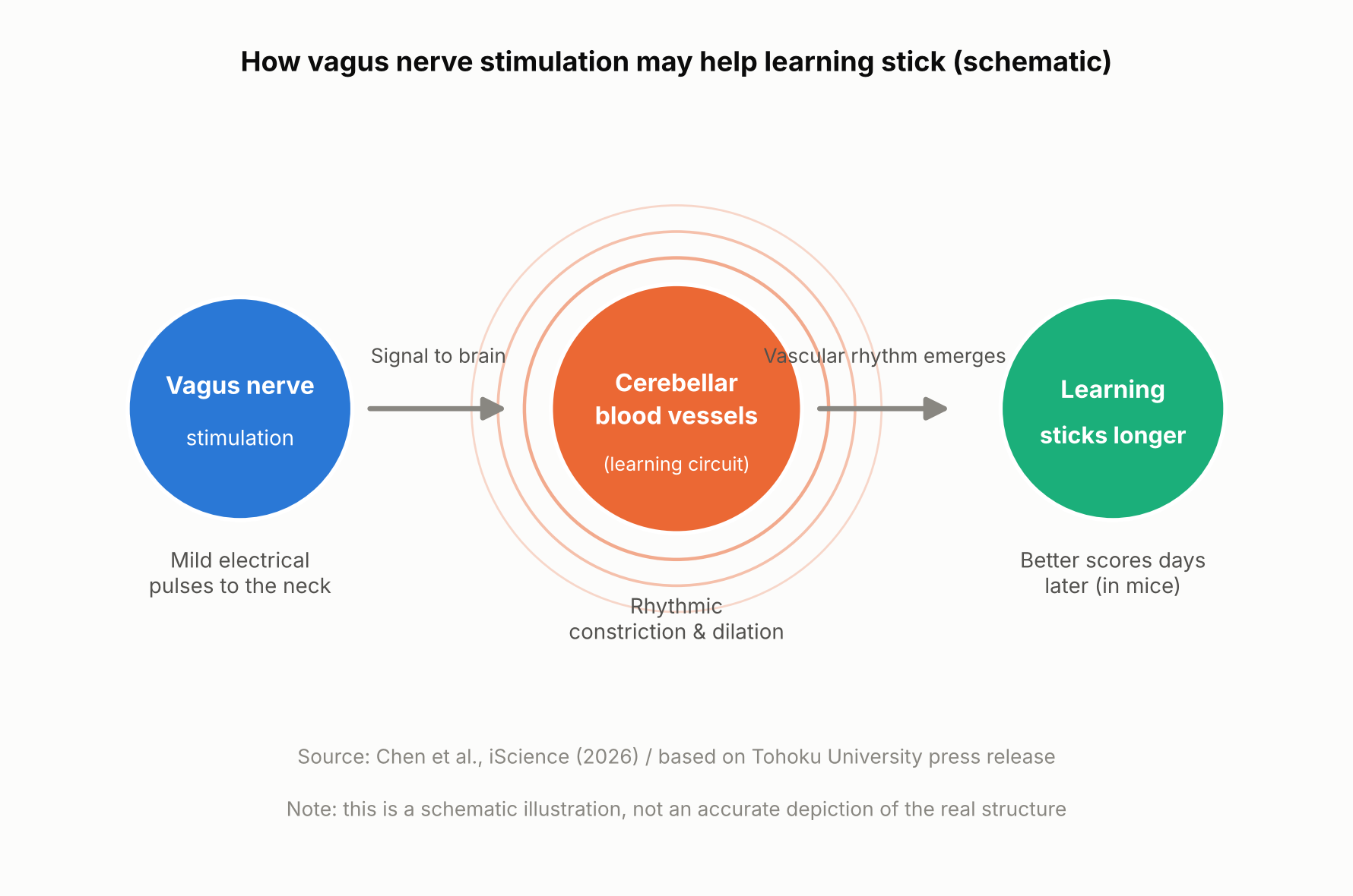
    Schematic showing how vagus nerve stimulation and a brain blood-volume rhythm relate to long-term learning
    How vagus nerve stimulation and a brain blood-volume rhythm relate to long-term learning (schematic illustration, not an accurate depiction of the real structure).

    The Mouse Experiment: Stimulation Came After Practice

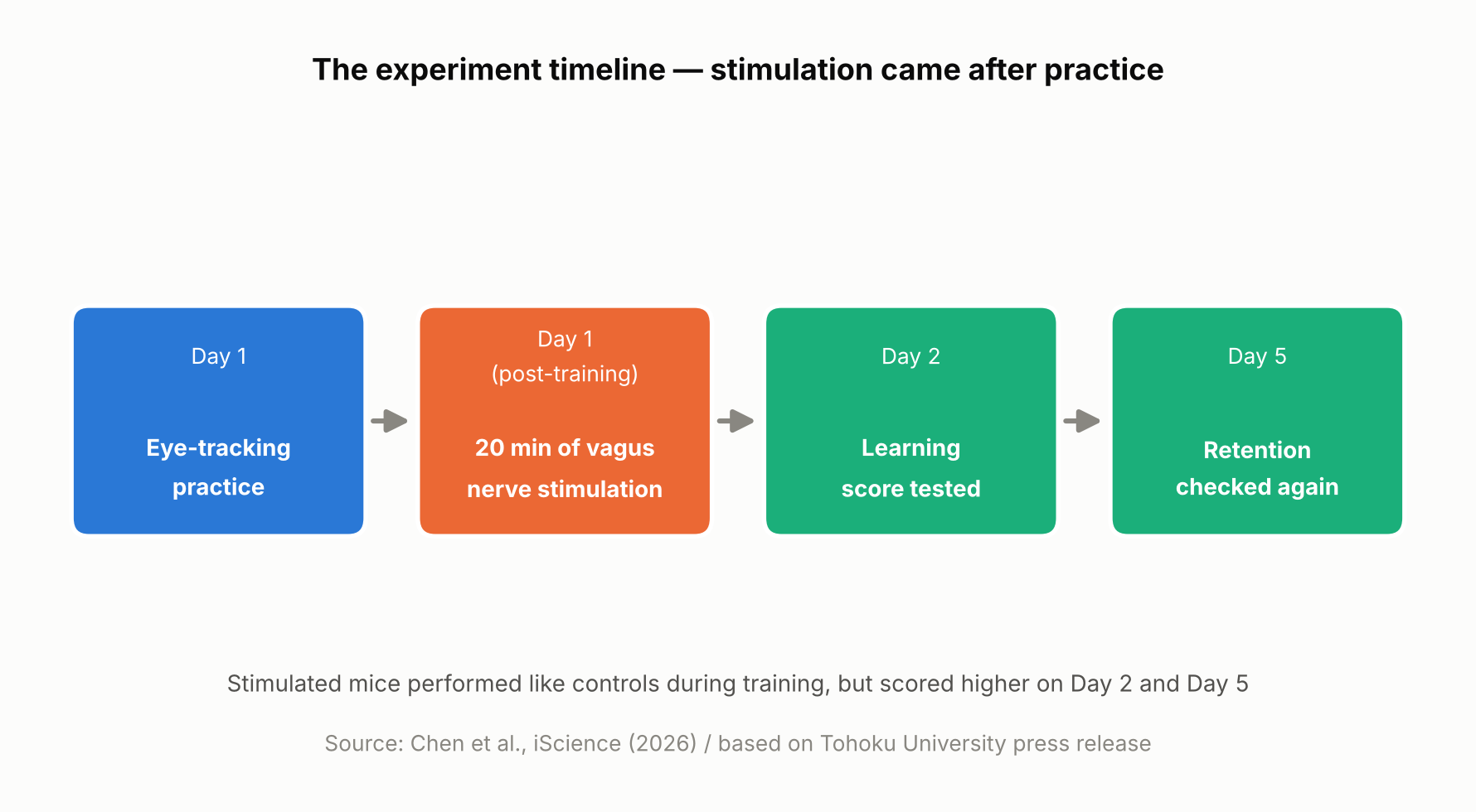
    A team at Tohoku University’s Graduate School of Life Sciences — Junyu U. Chen, Assistant Professor Yoko Ikoma, and Professor Ko Matsui — fitted a small cuff electrode around the left cervical vagus nerve in mice and delivered electrical stimulation. The mice practiced a task called the horizontal optokinetic response (HOKR): tracking a vertical striped pattern that moved back and forth in front of them with their eyes. This kind of task is known to depend heavily on the cerebellum, a brain region central to motor learning.

    On day one, the mice practiced for 15 minutes, four times, spaced an hour apart. Right after each day-one training session, mice in the stimulated group received 20 minutes of vagus nerve stimulation at one of two intensities, while control mice received none. Performance was then measured again on day two and day five.

    On day one itself, there was no difference between the stimulated and unstimulated mice. But by day two and especially day five, the stimulated mice showed higher scores, with the higher-intensity stimulation producing a visible benefit as early as day two and both intensities showing an advantage by day five. In other words, VNS didn’t directly boost performance during training — it appears to have improved later-day learning through some process that unfolded after training ended.

    Timeline of the mouse experiment, showing stimulation delivered after practice
    The experiment timeline. Based on Chen et al., iScience (2026) and the Tohoku University press release.

    Blood Volume Around the Cerebellum Fell Into Rhythm After Stimulation

    Using a light-based technique called fiber photometry, the researchers measured local blood-volume changes in tissue around the cerebellar flocculus. Each train of vagus nerve stimulation produced a two-step change: local blood volume briefly dipped, then — after a short delay — rose. This pattern is consistent with alternating contraction and dilation of blood vessels in that area.

    With repeated stimulation, this rise-and-fall pattern settled into a steady rhythm. The researchers think this rhythmic change in blood volume may be related to how the brain regulates local blood flow and its metabolic environment — but the study did not directly measure oxygen, ATP, or other metabolic markers, so exactly how the blood-volume rhythm might support learning is still unclear.

    Long-term learning is thought to involve various changes in the brain, including “synaptic plasticity” — changes in the connections and activity between neurons — which requires energy. The research team’s idea is that if the blood-volume rhythm is tied to how well-nourished brain tissue is, it could be helping support those changes, though this remains a hypothesis rather than something the study directly confirmed.

    Mice with larger blood-volume oscillations after stimulation tended to score higher on the learning test five days later

    A Bigger Rhythm, Better Long-Term Learning — but Only a Correlation So Far

    The most notable finding was a relationship between the magnitude of the blood-volume oscillations in the 20 minutes right after stimulation and performance five days later: mice with larger oscillations tended to score higher on day five. It’s worth being clear that this is a correlation, not proof that the blood-volume change caused the better learning.

    “The key point is that VNS was delivered only after training,” said Professor Matsui. “Our findings suggest that VNS may open a hidden window of opportunity for enhanced learning by making the brain environment more receptive to long-lasting change.” Lead author Junyu Chen added, “Our brains may be more strongly influenced by the body than we imagine. By tuning the brain’s metabolic environment, including rhythmic vascular movements, we may eventually unlock capacities that would otherwise remain latent.”

    What’s Still Unknown — and What Comes Next

    Based on this study alone, it’s not possible to say whether the blood-volume rhythm actually causes the learning improvement. Confirming that would require future experiments that directly manipulate blood-vessel or blood-volume activity and track the effect on learning. The researchers also did not directly measure metabolic molecules such as ATP, so the proposed mechanism remains a hypothesis.

    It’s also worth remembering that this was a mouse study using an invasive cuff electrode surgically placed around the cervical vagus nerve, delivering stimulation directly to the nerve rather than through the skin. Less invasive, ear-based stimulation methods are already being studied in humans for other purposes, but whether this mouse study’s results would carry over to humans — or to those non-invasive methods — can’t be determined from this result alone. The research team suggests that fine-tuning the timing and intensity of stimulation could eventually help support learning and rehabilitation, though that application remains a research goal rather than something available today.

    Sources: Tohoku University Graduate School of Life Sciences press release (August 26, 2026), Chen et al., iScience (published online August 25, 2026)

  • [Science] Scientists Build a Billion-Dalton Molecular Thread Longer Than a Cell — It Quadrupled Nerve-Cell Activity in the Lab

    [Science] Scientists Build a Billion-Dalton Molecular Thread Longer Than a Cell — It Quadrupled Nerve-Cell Activity in the Lab

    • A newly built, self-assembling material made nerve cells growing in a lab dish roughly four times more biologically active than usual, hinting that materials like it could one day help repair damaged nerves.
    • Chemists at Northwestern University found a new way to precisely control how long a certain kind of molecular thread grows, and how its positively and negatively charged segments are arranged along the way.
    • By molecular standards the resulting threads are enormous — roughly 100 times heavier than typical plastic-like polymers and bigger than even the largest proteins found in nature — even though they are built from many smaller molecules that click together on their own, rather than one molecule held together by ordinary chemical bonds.
    • So far the effect has only been shown in cells grown in a lab dish, not in animals or people, so any real treatment is still a long way off.

    Billion-Dalton Supramolecular Polymers: A Structure Big Enough to Rival a Cell

    A team led by the chemist Samuel I. Stupp at Northwestern University has built a new kind of giant structure, described in the journal Science on October 1, 2026. It belongs to a class of materials called supramolecular polymers. Rather than being a single molecule held together by strong chemical bonds the way an ordinary plastic is, it is made of many smaller molecules that click onto one another and hold themselves together more loosely, the way a line of magnets snaps together end to end. That looseness usually makes supramolecular polymers easy to assemble but hard to control, since there has been no reliable way to stop them from growing at a chosen point.

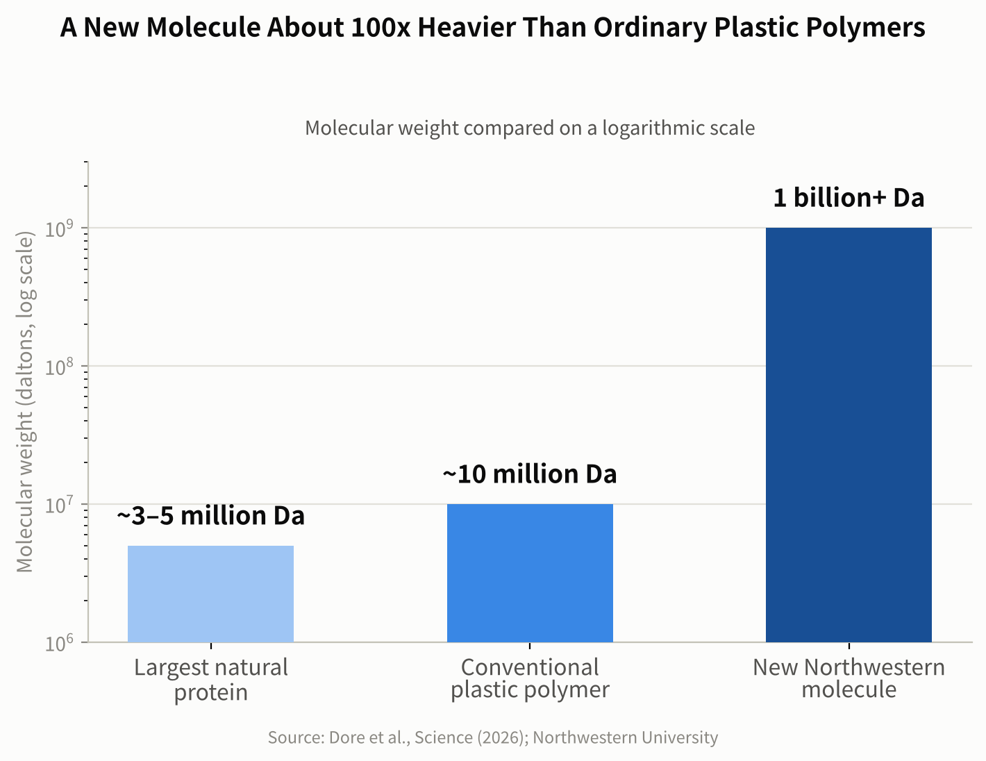
    Scientists weigh molecules in daltons, a unit named after the 19th-century chemist John Dalton, where one dalton is roughly the mass of a single hydrogen atom. Northwestern researchers point to common high-mass synthetic polymers used in materials science, which weigh around 10 million daltons, for comparison. Some of the largest proteins found in nature — the kind that give muscle tissue its structure — are in the roughly 3 to 5 million-dalton range. The new Northwestern assembly reaches a combined mass in the billions of daltons, around 100 times heavier than those conventional polymers, and its longest threads stretch farther than the width of a single living cell.

    Bar chart comparing molecular weight of a natural protein, a conventional polymer, and the new Northwestern assembly on a log scale
    Figure 1. Molecular weight on a log scale: a natural protein, a conventional polymer and the new billion-dalton supramolecular polymer. Source: data from Dore et al., Science (2026); Northwestern University

    Why Controlling the Length Was So Hard

    Because supramolecular polymers assemble themselves from many separate molecules rather than being built link by link under a chemist’s control, they have historically kept growing in a somewhat unpredictable way. That made it hard to control how long any given thread would grow, or where along its length particular features would end up. The Northwestern team solved this with a technique they call self-capping supramolecular polymerization: as the supply of available building-block molecules runs low, flexible regions at each end of the growing thread fold over on themselves, effectively capping the ends and stopping further growth.

    That trick let the researchers control two things at once: the overall length of each thread, and how positively and negatively charged segments are arranged along it. In a statement describing the work, Stupp called the result a breakthrough in materials design.

    “We discovered something we had never seen before in chemistry or in biology.”

    Precise Length and Charge Segmentation: The Charge Pattern Made the Difference

    Before arriving at their final design, the researchers tried several simpler charge patterns on neurons growing in a lab dish. Threads carrying only positive charge essentially killed the neurons; threads carrying only negative charge did almost nothing; and threads with positive and negative charge mixed randomly along their length produced no notable response either. Only one design worked well: a short positively charged segment flanked by longer negatively charged segments, arranged with precision rather than left to chance.

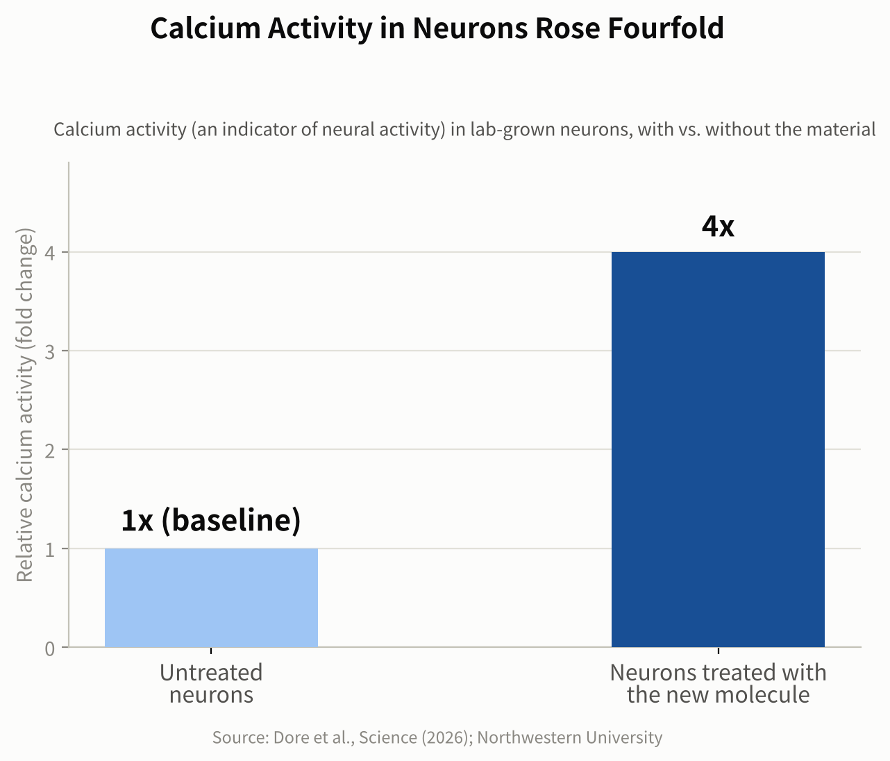
    To see how this winning design affected living neurons in culture, the researchers added it to cells growing in a lab dish and tracked them over 7 and 14 days. They used a technique called calcium imaging, which tracks what is known as calcium activity — a widely used indicator of how active a nerve cell is, since neurons briefly let calcium ions flow across their outer membrane as part of firing a signal. It is a standard, well-established way for researchers to gauge neural activity, though it is an indirect measure rather than a literal count of signals fired.

    Neurons treated with the winning design showed overall calcium activity roughly four times greater than untreated neurons after seven days, and had formed more synapses by 14 days. The researchers propose that the precise charge pattern works by shaping how the material interacts with the neurons’ outer membrane and with proteins around it, though they describe this as a proposed mechanism rather than a fully confirmed one. Notably, the effect appeared without adding any separate drug, growth factor, or other biological signaling molecule — the geometry of the charge pattern alone was enough to measurably change how the cells behaved.

    That matters because nerve cells are known to rely on their electrical activity, along with other cues, to grow new branches and form synapses, the junctions where one neuron passes a signal to the next. Materials associated with more active, better-connected neurons are therefore of particular interest for regenerative medicine, the branch of science focused on helping the body rebuild tissue it has lost or damaged, including nerve tissue after an injury. The researchers describe the link between an entirely synthetic, self-assembling material and this kind of change in living cells as the most surprising part of the result.

    Bar chart showing neurons treated with the new material had four times the calcium activity of untreated neurons
    Figure 2. Neurons treated with the new material showed about four times the calcium activity of untreated neurons. Source: data from Dore et al., Science (2026); Northwestern University

    From a Lab Dish to a Possible Therapy

    So far this particular design has only been tested on neurons growing in a dish, not in animals or people, so any medical use is still likely years away. Lab-dish results are a long way from a treatment: cells in a dish do not have to survive inside a living body, fight off an immune response, or integrate with surrounding tissue the way they would after an actual injury. The Stupp lab has, however, spent years designing self-assembling materials aimed at nerve repair and tissue regeneration, and the researchers see this new level of precision as a step toward building more reliable, tunable materials for that purpose. A logical next stage would be testing whether the material remains safe and effective in animal models of nerve damage, before any human study could even be considered.

    The achievement also matters well beyond medicine. Being able to build a giant synthetic structure with a chosen length and a precisely controlled charge pattern opens the door to new materials with tunable mechanical or electrical behavior, useful in fields far from biology too. More broadly, it shows how self-assembly can be used to control a material’s properties over distances far larger than a single molecule — a step toward manufacturing matter with a level of precision that has mostly been limited, until now, to much smaller scales.

    Sources: Northwestern Now, EurekAlert!

    Paper: Dore, M. et al., “Precise length and charge segmentation of billion-dalton supramolecular polymers”, Science (2026).

    Related: new to the topic? Start with What Is a Supramolecular Polymer? A Simple Guide to How It Works.

  • [Science] The Instant You Decide, Your Brain Re-Files the Options — MIT Neuroscientists Find a Flexible Code That Could Inform AI Design

    [Science] The Instant You Decide, Your Brain Re-Files the Options — MIT Neuroscientists Find a Flexible Code That Could Inform AI Design

    Every time you choose between two options — which job offer to accept, which product to buy — your brain does more than simply retrieve what each one was worth. A new MIT study shows that the instant you make that choice, the neural code representing those options in part of your brain reorganizes itself, shifting from grouping information by the order you saw it to grouping it by whether you picked it. The finding, published online in the journal iScience in September 2026, offers a rare, cell-by-cell look at how flexible biological memory can be — and it gives researchers working on adaptive AI a biological case study worth studying, even though the experiment itself did not involve any AI system.

    Why Scientists Wanted to Watch the Brain Mid-Decision

    Researchers have long known that the prefrontal cortex, the region of the brain just behind the forehead, plays a central role in weighing options and making choices. Many of its neurons also show “mixed selectivity,” meaning a single cell’s activity can shift depending on a combination of task variables rather than tracking one fixed thing. What earlier work left unresolved was how the brain avoids mixing up two options that were evaluated only moments apart, especially once one has been chosen and the other set aside. A team led by MIT graduate student Huidi Li and senior author Earl K. Miller, a professor at MIT’s Picower Institute for Learning and Memory, set out to track that process neuron by neuron rather than assume an answer. The team, which also included Nikolaos Chrysanthidis, Scott Brincat, and Jonas Rose, published its results in iScience; MIT publicized the findings on September 22.

    Watching Hundreds of Neurons Decide, in Real Time

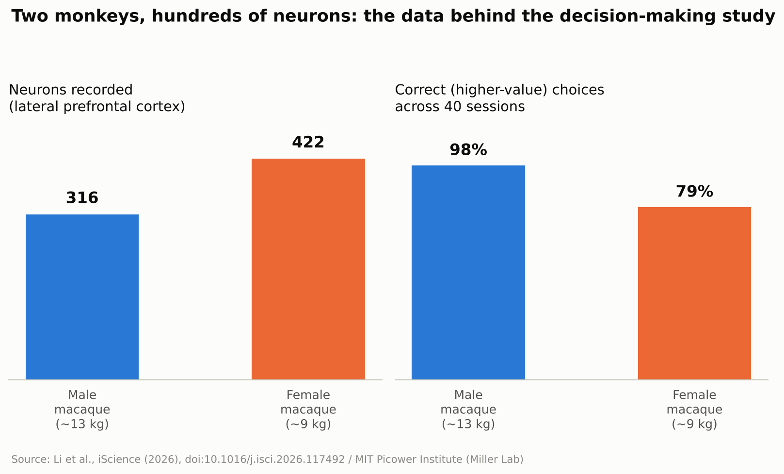
    To find out, the researchers trained two macaque monkeys — a roughly 13-kilogram male and a 9-kilogram female — to play a simple reward game. A white square flashed briefly at one of four spots on a screen, followed by a colored ring that signaled how many drops of juice that option was worth, anywhere from one to thirteen. The same sequence then repeated for a second target at a different screen location. Once both options had been shown, the monkey looked toward its preferred target and received the corresponding juice reward. The male selected the higher-value option on about 98 percent of trials; the female, on about 79 percent. While the animals made their choices, the team recorded the electrical activity of 316 neurons in the male’s lateral prefrontal cortex — the side portion of the prefrontal cortex — and 422 in the female’s, across 40 recording sessions. Rather than tracking single cells in isolation, the researchers analyzed the activity of the whole recorded population as points moving through a high-dimensional mathematical space, a technique known as neural subspace analysis: when enough neurons fire in a similar combined pattern, that pattern occupies its own region, or “subspace,” within that space, and two subspaces that barely overlap are described as close to orthogonal, or mathematically independent of each other.

    Bar chart showing neurons recorded and choice accuracy for two macaques in the study
    Source: Created from data in Li et al., iScience (2026)

    Before the Choice: Order. After the Choice: Outcome.

    Seen through that lens, the result is a clear shift in organization. Before a choice was made, the subspaces representing the first-shown and second-shown targets were kept largely separate, organized around presentation order. Once the monkey chose, the picture changed: the subspaces reorganized around the decision itself, with the chosen and unchosen options rotating into distinct, close-to-orthogonal subspaces regardless of the order in which they had originally appeared. In other words, the brain’s filing system for “what I saw first and second” gave way to a new one built around “what I picked and what I didn’t.” The researchers suggest this reorganization helps downstream brain regions read out the decision efficiently, since they could rely on a consistent format rather than track which position each option had occupied.

    “The brain doesn’t just hold information statically,” Li said.

    No Single Neuron “Owns” a Decision

    A second notable result was how that reorganization happened at the level of individual cells. The researchers found no fixed set of neurons permanently dedicated to representing “the chosen option” or “the rejected one.” Instead, individual neurons shifted which ensemble they contributed to from one trial to the next, joining a different, temporary coalition of cells depending on what the task required at that moment. Researchers in the field generally see this kind of flexible, overlapping coding as a way for a limited number of neurons to represent many different combinations of information, rather than requiring a dedicated cell type for every possible scenario — though this study did not directly measure how much representational capacity that flexibility adds.

    Why This Matters Beyond the Lab

    The study did not test any artificial intelligence system, and the researchers were not trying to build one. Still, the result speaks to a question AI researchers care about: how can a learning system keep updating what it represents without losing track of information it already holds? Many AI systems, once trained, represent what they have learned in ways that stay largely fixed afterward, which is part of why training them further on new material can degrade old knowledge, a problem researchers call catastrophic forgetting. The MIT findings do not explain or solve that problem, but the underlying idea — a biological circuit reorganizing its internal representations on the fly to match what a task demands at each stage — is the kind of mechanism that researchers in continual learning and brain-inspired, or neuromorphic, computing study for design ideas. It is also a small, concrete answer to a more everyday question: the researchers found that even after a choice was made, the unchosen option remained represented in its own distinct subspace rather than disappearing from the recorded activity. MIT’s own description of the work suggested this could be important for later crediting an outcome to the option that produced it, which may help the brain learn from an alternative it did not select — though that specific learning process was not directly tested in this experiment.

    What’s Still Unknown

    The authors are careful to flag the limits of the work. Only two animals took part, and the task involved a simple, sequential choice between two options with clearly different, discrete reward values — closer to comparing two job offers one after the other than to scanning a crowded menu of possibilities at once. Whether the same kind of reorganization occurs when values vary continuously, when there are more than two options on the table, or in human decision-making specifically, remains an open question the team says future studies will need to address. Macaque and human prefrontal cortex share many organizational similarities, which is why monkey studies like this one are routinely used to generate hypotheses about human cognition, but confirming that the same reorganization happens in people would require a separate study in humans. It is also worth remembering that this was a recording study: it shows that representations change alongside decisions, not that the reorganization itself is what causes a choice to happen. As with any single study, independent replication will matter before the finding is considered settled.

    For now, the study offers a concrete, carefully measured answer to a question that sounds almost philosophical: what happens in your head in the instant after you decide? According to this research, at least in one brain region of two monkeys performing a simple task, the answer is that the brain quietly rebuilds how it was organizing two competing options around the choice that was just made.

    Sources: MIT News, iScience (Li et al., 2026), EurekAlert!