Tag: Learning

  • [Science] Why Letting AI Help Can Erode Your Grit, Berkeley Study Finds

    [Science] Why Letting AI Help Can Erode Your Grit, Berkeley Study Finds

    A chatbot that hands you the answer can make a hard problem feel easy — until the chatbot is gone. A new study from researchers at five universities found that people who solved problems with an AI assistant performed worse on the next problems, once that assistant was taken away, than people who had worked through the very same problems alone from the start. The gap showed up after roughly 10 to 15 minutes of AI-assisted problem-solving, and it held across three separate experiments involving 1,222 recruited participants — a pattern the authors say raises real questions about whether leaning on AI for answers can undermine people’s ability, or willingness, to solve problems on their own.

    Three Experiments, One Pattern

    The team — Grace Liu, Brian Christian, Tsvetomira Dumbalska, Michiel A. Bakker and Rachit Dubey, drawing on UC Berkeley’s Center for Human-Compatible AI along with Carnegie Mellon University, MIT, the University of Oxford and UCLA — ran three randomized controlled trials totaling 1,222 recruited participants (307, 585 and 168 after some participants were excluded under the researchers’ prespecified criteria). In the first experiment, 354 people were recruited to solve 12 basic fraction problems; half worked alone throughout, while the other half could ask a ChatGPT assistant, built on GPT-5, for help — before everyone, AI group included, had to solve three further problems entirely on their own. A second experiment repeated that design with 667 people, and a third, with 201 participants, swapped the math problems for SAT-style reading comprehension questions.

    In every version of the study, the early pattern looked encouraging: while they still had access to the AI, the assisted group solved problems faster and scored higher than the group working alone — exactly what you would expect from a capable assistant. The twist came in the second half of each session, once the AI was removed and everyone, including the AI group, had to finish the rest of the problems unaided.

    The Numbers Behind the Finding

    Once AI access ended, the previously assisted group fell behind the group that had never used it, and the gap was consistent across all three experiments. In the first fraction experiment, the former AI group answered an average of 57% of the three final problems correctly, versus 73% for the no-AI group — a statistically significant difference (p < 0.001). The larger replication of the same design found a smaller but still reliable gap: 71% versus 77% (p = 0.02). In the reading-comprehension experiment, the difference widened again, to 76% versus 89% (p = 0.007).
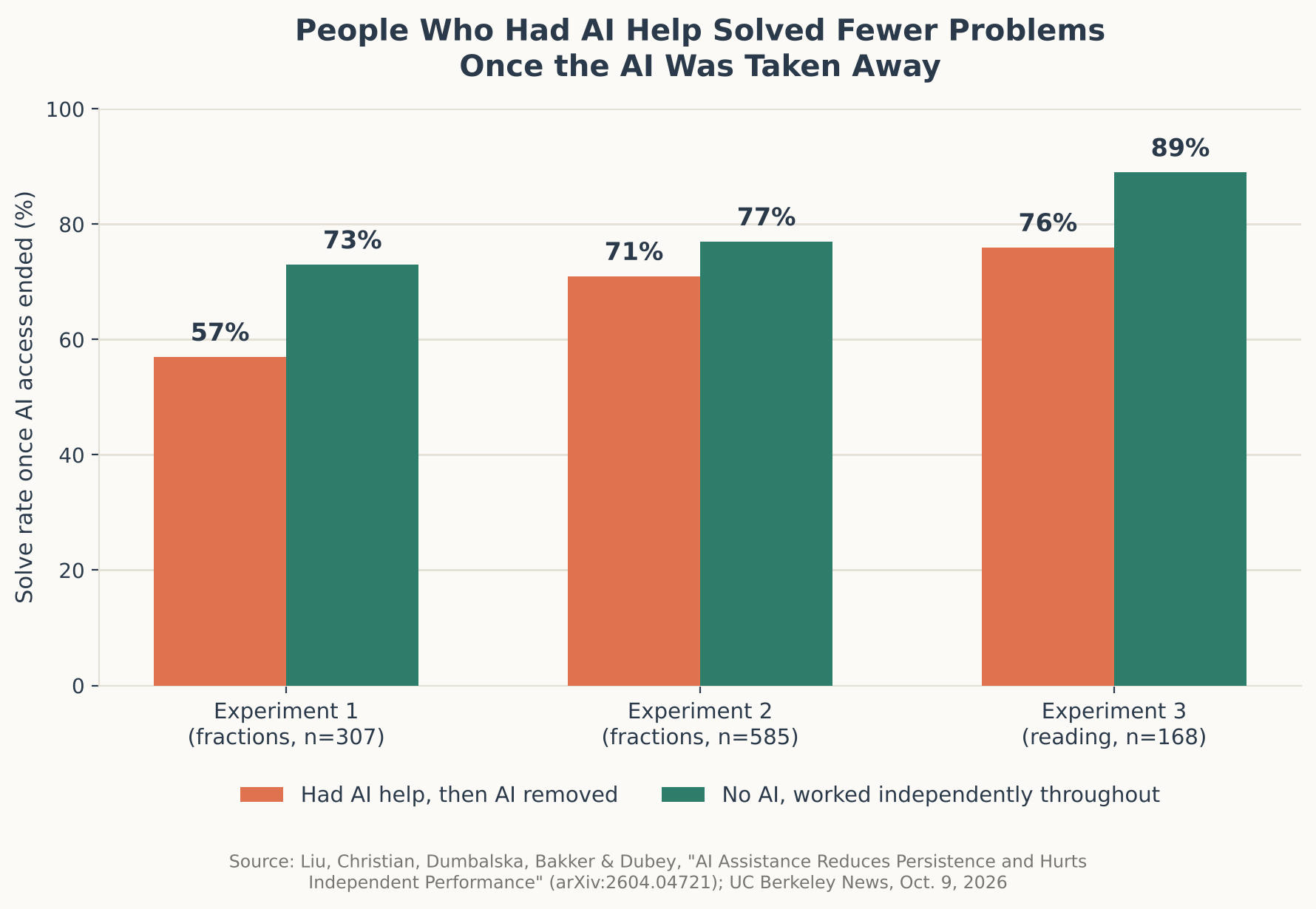
    Bar chart comparing solve rates after AI was removed across three experiments
    Source: data from Liu, Christian, Dumbalska, Bakker & Dubey (arXiv:2604.04721) and UC Berkeley News

    Giving up altogether followed a similar trend. In the first experiment, 20% of the former AI group skipped a problem rather than attempt it, compared with 11% of the no-AI group. The pattern was weaker in the larger replication (10% versus 7%, not statistically significant) but reappeared clearly in the reading task, where 8% of the AI group skipped questions versus just 1% of the control group. A closer look at how participants said they had actually used the AI while it was available sharpened the picture further, though this part of the analysis was observational rather than randomized, since people were not assigned to copy answers or ask for hints: those who reported simply copying the AI’s finished answers fared worst afterward, averaging just 65% correct on their own, while those who said they used the AI mainly for hints showed no statistically significant gap from people who never touched AI at all.

    Participants were randomly assigned to the AI or no-AI condition, and the larger replication added a pretest to check the groups further: accuracy and the tendency to skip a problem did not differ significantly between groups beforehand, which strengthens the case that the later gap reflects the AI condition itself rather than pre-existing differences between participants. In that same larger replication, participants were also asked how they had actually used the AI while it was available. A small slice, about 12%, said they had barely used it despite having access, and this group performed about as well afterward as people who never had AI access at all — reinforcing the idea that it is the degree of reliance on AI, not mere availability of it, that predicts the later drop-off.

    “It’s a striking finding, but in a way it supplies ammunition for a story that a lot of us kind of feel in our gut.” — Brian Christian, UC Berkeley

    What Might Explain the Pattern

    The authors propose two explanations, while cautioning that their experiments were not designed to prove which one is doing the work. The first is a kind of expectation mismatch: once someone gets used to near-instant answers, grinding through a problem by hand can suddenly feel disproportionately slow, which may make people more willing to give up rather than push through. The second is the loss of what the researchers call “productive struggle” — the idea that difficulty itself is not simply an obstacle to route around, but part of how people practice and strengthen the skills a problem requires. Bypass that effort often enough, the argument goes, and there may be fewer chances for those skills to get built in the first place.

    It’s a different mechanism, but this study joins a related question this site has covered before: Georgetown University researchers recently found that AI-generated summaries of events can distort people’s memory of what actually happened. That study is about memory rather than problem-solving, but together the two point to a broader pattern worth watching — handing a mental task to AI can carry a hidden cost to accuracy or independent ability, even when it feels like pure convenience in the moment.

    “Not Just Fractions and SAT Problems”

    Christian has been blunt about how far he thinks the implications reach beyond a psychology lab. “This is not a story about fractions and SAT problems,” he said in comments accompanying the study’s release. “This is something that is happening to human knowledge and human expertise.” He described the irony directly: widely used AI assistants, he said, are “often helping us in ways that are kind of unhelpful” — lifting performance on the task directly in front of us while, the data suggest, working against the independent competence we will need the next time no AI is around.

    The caveats matter here. The study measured performance minutes after AI access ended, not months or years later, so it cannot say how long this effect lasts or whether frequent users eventually adapt and recover. The breakdown by how people used the AI — copying answers versus asking for hints — was observational rather than randomized, so it shows an association rather than definitive proof of cause and effect. And all three experiments involved short, one-off tasks completed by paid online participants, a different setting from a student using AI across a semester of coursework or an employee weaving it into daily work.

    What This Means for Using AI

    None of this is an argument for avoiding AI assistance altogether — the same data show it clearly boosted short-term performance while it was available, and for many real-world tasks, getting the right answer quickly is exactly the point. The more useful takeaway from this one study may be about how AI gets used rather than whether: in the self-reported usage data, the steepest drop-off belonged to people who let the AI hand them a finished answer, while those who said they used it mainly for hints or partial guidance looked much closer to the group that never used AI at all. Treating a chatbot more like a tutor that nudges you toward a solution, rather than a vending machine that dispenses the finished one, looks like a reasonable bet for keeping your own problem-solving sharp — though it’s one study, in a narrow setting, and confirming how far that advice travels beyond fractions and reading quizzes will take further research.

    Sources: UC Berkeley News, arXiv:2604.04721, “AI Assistance Reduces Persistence and Hurts Independent Performance”
  • [Science] Stimulating the Vagus Nerve After Practice Helps Mice Learn Better Days Later — Tohoku University Finds a Link to Rhythmic Changes in Brain Blood Volume

    [Science] Stimulating the Vagus Nerve After Practice Helps Mice Learn Better Days Later — Tohoku University Finds a Link to Rhythmic Changes in Brain Blood Volume

    • Applying electrical stimulation to the “vagus nerve” on the left side of the neck right after practicing something new may make mice better at it days later, a new study suggests.
    • If a similar effect holds in humans, it could eventually point toward new ways to support motor learning or rehabilitation.
    • Tohoku University researchers stimulated the vagus nerve while tracking local blood-volume changes in the brain, and found that repeated stimulation produced rhythmic fluctuations in local blood volume.
    • Mice with larger blood-volume oscillations after stimulation tended to score higher on a learning test five days later. This is a mouse study, though, and it’s not yet known whether the same effect would occur in humans.

    The Vagus Nerve — a Key Line Between Brain and Body

    The vagus nerve is a major nerve that runs from the brainstem through the neck, chest, and abdomen, carrying signals between the brain and organs such as the heart, lungs, and digestive tract. It’s one of the main nerves connecting the brain and internal organs in both directions: it carries signals about the body’s internal state — such as heart rate, breathing, and digestion — up to the brain, while also carrying signals from the brain back down to those organs.

    A treatment that stimulates this nerve from the outside with electrical pulses is called “vagus nerve stimulation,” or VNS. Using an implanted device, it’s an established therapy for epilepsy that doesn’t respond well enough to medication. In recent years, researchers have also been studying a non-invasive approach that stimulates part of the ear through the skin — though that’s a different method from the one used in this mouse study. Exactly what VNS changes inside the brain has remained unclear.

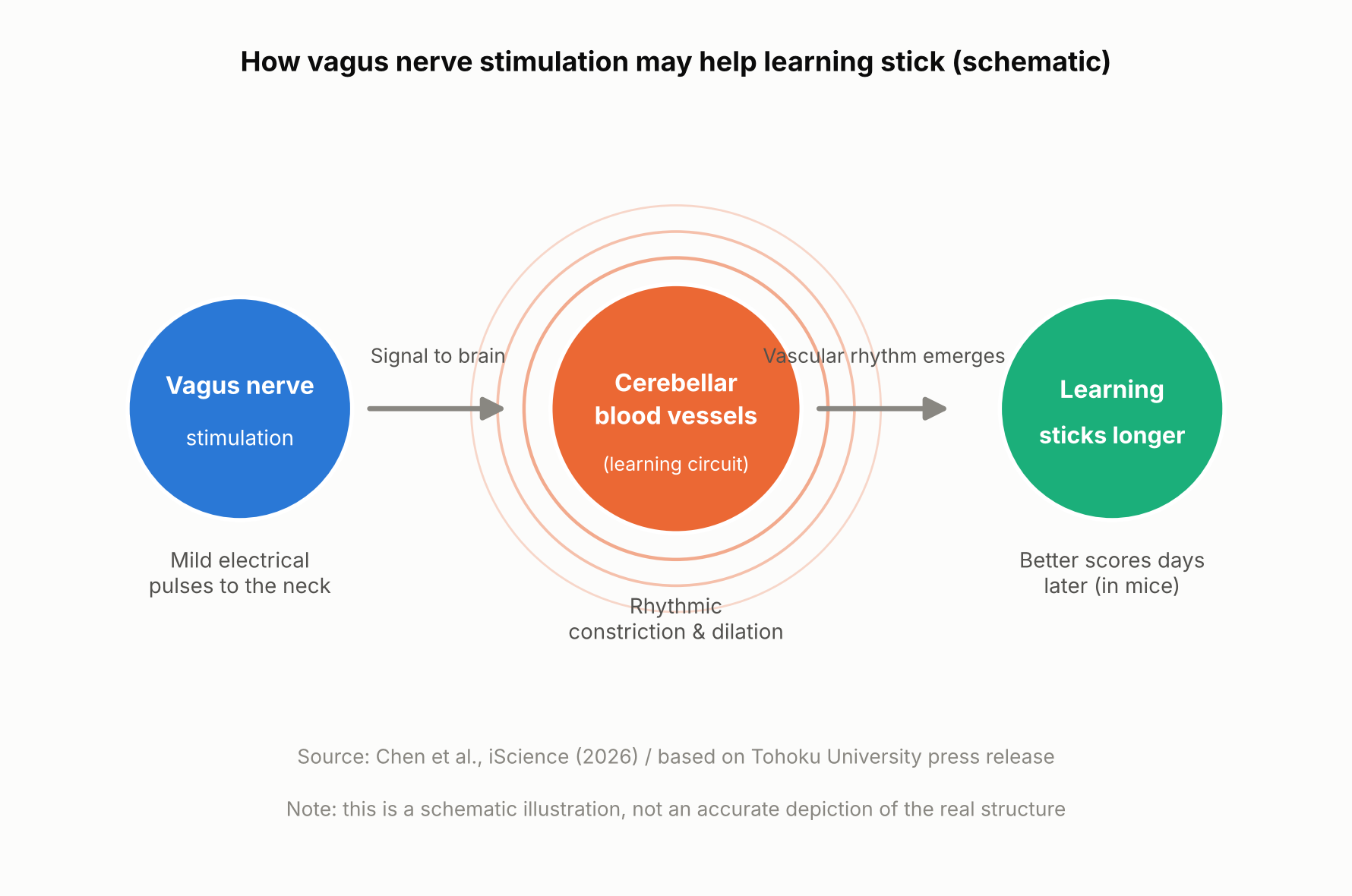
    Schematic showing how vagus nerve stimulation and a brain blood-volume rhythm relate to long-term learning
    How vagus nerve stimulation and a brain blood-volume rhythm relate to long-term learning (schematic illustration, not an accurate depiction of the real structure).

    The Mouse Experiment: Stimulation Came After Practice

    A team at Tohoku University’s Graduate School of Life Sciences — Junyu U. Chen, Assistant Professor Yoko Ikoma, and Professor Ko Matsui — fitted a small cuff electrode around the left cervical vagus nerve in mice and delivered electrical stimulation. The mice practiced a task called the horizontal optokinetic response (HOKR): tracking a vertical striped pattern that moved back and forth in front of them with their eyes. This kind of task is known to depend heavily on the cerebellum, a brain region central to motor learning.

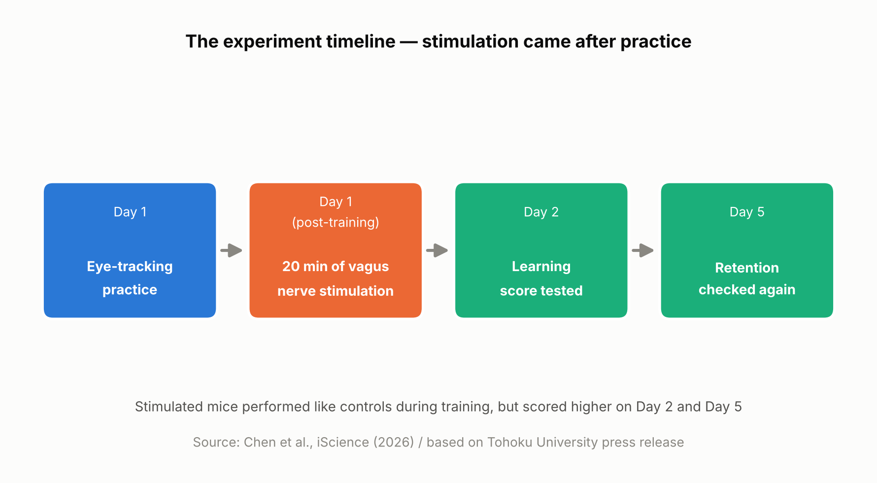
    On day one, the mice practiced for 15 minutes, four times, spaced an hour apart. Right after each day-one training session, mice in the stimulated group received 20 minutes of vagus nerve stimulation at one of two intensities, while control mice received none. Performance was then measured again on day two and day five.

    On day one itself, there was no difference between the stimulated and unstimulated mice. But by day two and especially day five, the stimulated mice showed higher scores, with the higher-intensity stimulation producing a visible benefit as early as day two and both intensities showing an advantage by day five. In other words, VNS didn’t directly boost performance during training — it appears to have improved later-day learning through some process that unfolded after training ended.

    Timeline of the mouse experiment, showing stimulation delivered after practice
    The experiment timeline. Based on Chen et al., iScience (2026) and the Tohoku University press release.

    Blood Volume Around the Cerebellum Fell Into Rhythm After Stimulation

    Using a light-based technique called fiber photometry, the researchers measured local blood-volume changes in tissue around the cerebellar flocculus. Each train of vagus nerve stimulation produced a two-step change: local blood volume briefly dipped, then — after a short delay — rose. This pattern is consistent with alternating contraction and dilation of blood vessels in that area.

    With repeated stimulation, this rise-and-fall pattern settled into a steady rhythm. The researchers think this rhythmic change in blood volume may be related to how the brain regulates local blood flow and its metabolic environment — but the study did not directly measure oxygen, ATP, or other metabolic markers, so exactly how the blood-volume rhythm might support learning is still unclear.

    Long-term learning is thought to involve various changes in the brain, including “synaptic plasticity” — changes in the connections and activity between neurons — which requires energy. The research team’s idea is that if the blood-volume rhythm is tied to how well-nourished brain tissue is, it could be helping support those changes, though this remains a hypothesis rather than something the study directly confirmed.

    Mice with larger blood-volume oscillations after stimulation tended to score higher on the learning test five days later

    A Bigger Rhythm, Better Long-Term Learning — but Only a Correlation So Far

    The most notable finding was a relationship between the magnitude of the blood-volume oscillations in the 20 minutes right after stimulation and performance five days later: mice with larger oscillations tended to score higher on day five. It’s worth being clear that this is a correlation, not proof that the blood-volume change caused the better learning.

    “The key point is that VNS was delivered only after training,” said Professor Matsui. “Our findings suggest that VNS may open a hidden window of opportunity for enhanced learning by making the brain environment more receptive to long-lasting change.” Lead author Junyu Chen added, “Our brains may be more strongly influenced by the body than we imagine. By tuning the brain’s metabolic environment, including rhythmic vascular movements, we may eventually unlock capacities that would otherwise remain latent.”

    What’s Still Unknown — and What Comes Next

    Based on this study alone, it’s not possible to say whether the blood-volume rhythm actually causes the learning improvement. Confirming that would require future experiments that directly manipulate blood-vessel or blood-volume activity and track the effect on learning. The researchers also did not directly measure metabolic molecules such as ATP, so the proposed mechanism remains a hypothesis.

    It’s also worth remembering that this was a mouse study using an invasive cuff electrode surgically placed around the cervical vagus nerve, delivering stimulation directly to the nerve rather than through the skin. Less invasive, ear-based stimulation methods are already being studied in humans for other purposes, but whether this mouse study’s results would carry over to humans — or to those non-invasive methods — can’t be determined from this result alone. The research team suggests that fine-tuning the timing and intensity of stimulation could eventually help support learning and rehabilitation, though that application remains a research goal rather than something available today.

    Sources: Tohoku University Graduate School of Life Sciences press release (August 26, 2026), Chen et al., iScience (published online August 25, 2026)

  • [Science] The Instant You Decide, Your Brain Re-Files the Options — MIT Neuroscientists Find a Flexible Code That Could Inform AI Design

    [Science] The Instant You Decide, Your Brain Re-Files the Options — MIT Neuroscientists Find a Flexible Code That Could Inform AI Design

    Every time you choose between two options — which job offer to accept, which product to buy — your brain does more than simply retrieve what each one was worth. A new MIT study shows that the instant you make that choice, the neural code representing those options in part of your brain reorganizes itself, shifting from grouping information by the order you saw it to grouping it by whether you picked it. The finding, published online in the journal iScience in September 2026, offers a rare, cell-by-cell look at how flexible biological memory can be — and it gives researchers working on adaptive AI a biological case study worth studying, even though the experiment itself did not involve any AI system.

    Why Scientists Wanted to Watch the Brain Mid-Decision

    Researchers have long known that the prefrontal cortex, the region of the brain just behind the forehead, plays a central role in weighing options and making choices. Many of its neurons also show “mixed selectivity,” meaning a single cell’s activity can shift depending on a combination of task variables rather than tracking one fixed thing. What earlier work left unresolved was how the brain avoids mixing up two options that were evaluated only moments apart, especially once one has been chosen and the other set aside. A team led by MIT graduate student Huidi Li and senior author Earl K. Miller, a professor at MIT’s Picower Institute for Learning and Memory, set out to track that process neuron by neuron rather than assume an answer. The team, which also included Nikolaos Chrysanthidis, Scott Brincat, and Jonas Rose, published its results in iScience; MIT publicized the findings on September 22.

    Watching Hundreds of Neurons Decide, in Real Time

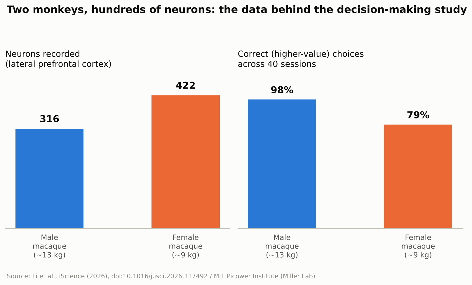
    To find out, the researchers trained two macaque monkeys — a roughly 13-kilogram male and a 9-kilogram female — to play a simple reward game. A white square flashed briefly at one of four spots on a screen, followed by a colored ring that signaled how many drops of juice that option was worth, anywhere from one to thirteen. The same sequence then repeated for a second target at a different screen location. Once both options had been shown, the monkey looked toward its preferred target and received the corresponding juice reward. The male selected the higher-value option on about 98 percent of trials; the female, on about 79 percent. While the animals made their choices, the team recorded the electrical activity of 316 neurons in the male’s lateral prefrontal cortex — the side portion of the prefrontal cortex — and 422 in the female’s, across 40 recording sessions. Rather than tracking single cells in isolation, the researchers analyzed the activity of the whole recorded population as points moving through a high-dimensional mathematical space, a technique known as neural subspace analysis: when enough neurons fire in a similar combined pattern, that pattern occupies its own region, or “subspace,” within that space, and two subspaces that barely overlap are described as close to orthogonal, or mathematically independent of each other.

    Bar chart showing neurons recorded and choice accuracy for two macaques in the study
    Source: Created from data in Li et al., iScience (2026)

    Before the Choice: Order. After the Choice: Outcome.

    Seen through that lens, the result is a clear shift in organization. Before a choice was made, the subspaces representing the first-shown and second-shown targets were kept largely separate, organized around presentation order. Once the monkey chose, the picture changed: the subspaces reorganized around the decision itself, with the chosen and unchosen options rotating into distinct, close-to-orthogonal subspaces regardless of the order in which they had originally appeared. In other words, the brain’s filing system for “what I saw first and second” gave way to a new one built around “what I picked and what I didn’t.” The researchers suggest this reorganization helps downstream brain regions read out the decision efficiently, since they could rely on a consistent format rather than track which position each option had occupied.

    “The brain doesn’t just hold information statically,” Li said.

    No Single Neuron “Owns” a Decision

    A second notable result was how that reorganization happened at the level of individual cells. The researchers found no fixed set of neurons permanently dedicated to representing “the chosen option” or “the rejected one.” Instead, individual neurons shifted which ensemble they contributed to from one trial to the next, joining a different, temporary coalition of cells depending on what the task required at that moment. Researchers in the field generally see this kind of flexible, overlapping coding as a way for a limited number of neurons to represent many different combinations of information, rather than requiring a dedicated cell type for every possible scenario — though this study did not directly measure how much representational capacity that flexibility adds.

    Why This Matters Beyond the Lab

    The study did not test any artificial intelligence system, and the researchers were not trying to build one. Still, the result speaks to a question AI researchers care about: how can a learning system keep updating what it represents without losing track of information it already holds? Many AI systems, once trained, represent what they have learned in ways that stay largely fixed afterward, which is part of why training them further on new material can degrade old knowledge, a problem researchers call catastrophic forgetting. The MIT findings do not explain or solve that problem, but the underlying idea — a biological circuit reorganizing its internal representations on the fly to match what a task demands at each stage — is the kind of mechanism that researchers in continual learning and brain-inspired, or neuromorphic, computing study for design ideas. It is also a small, concrete answer to a more everyday question: the researchers found that even after a choice was made, the unchosen option remained represented in its own distinct subspace rather than disappearing from the recorded activity. MIT’s own description of the work suggested this could be important for later crediting an outcome to the option that produced it, which may help the brain learn from an alternative it did not select — though that specific learning process was not directly tested in this experiment.

    What’s Still Unknown

    The authors are careful to flag the limits of the work. Only two animals took part, and the task involved a simple, sequential choice between two options with clearly different, discrete reward values — closer to comparing two job offers one after the other than to scanning a crowded menu of possibilities at once. Whether the same kind of reorganization occurs when values vary continuously, when there are more than two options on the table, or in human decision-making specifically, remains an open question the team says future studies will need to address. Macaque and human prefrontal cortex share many organizational similarities, which is why monkey studies like this one are routinely used to generate hypotheses about human cognition, but confirming that the same reorganization happens in people would require a separate study in humans. It is also worth remembering that this was a recording study: it shows that representations change alongside decisions, not that the reorganization itself is what causes a choice to happen. As with any single study, independent replication will matter before the finding is considered settled.

    For now, the study offers a concrete, carefully measured answer to a question that sounds almost philosophical: what happens in your head in the instant after you decide? According to this research, at least in one brain region of two monkeys performing a simple task, the answer is that the brain quietly rebuilds how it was organizing two competing options around the choice that was just made.

    Sources: MIT News, iScience (Li et al., 2026), EurekAlert!