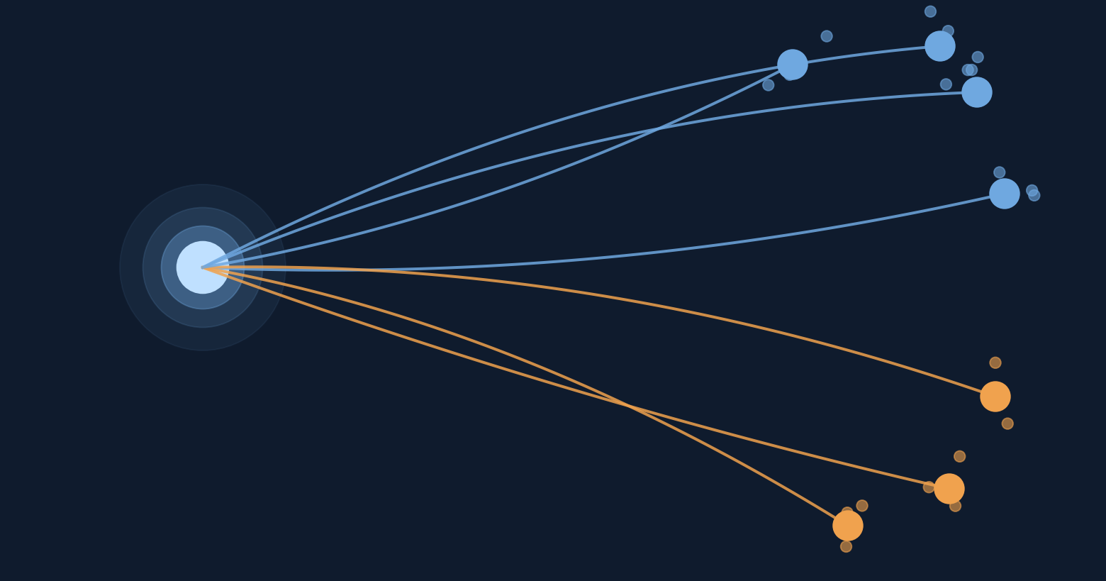
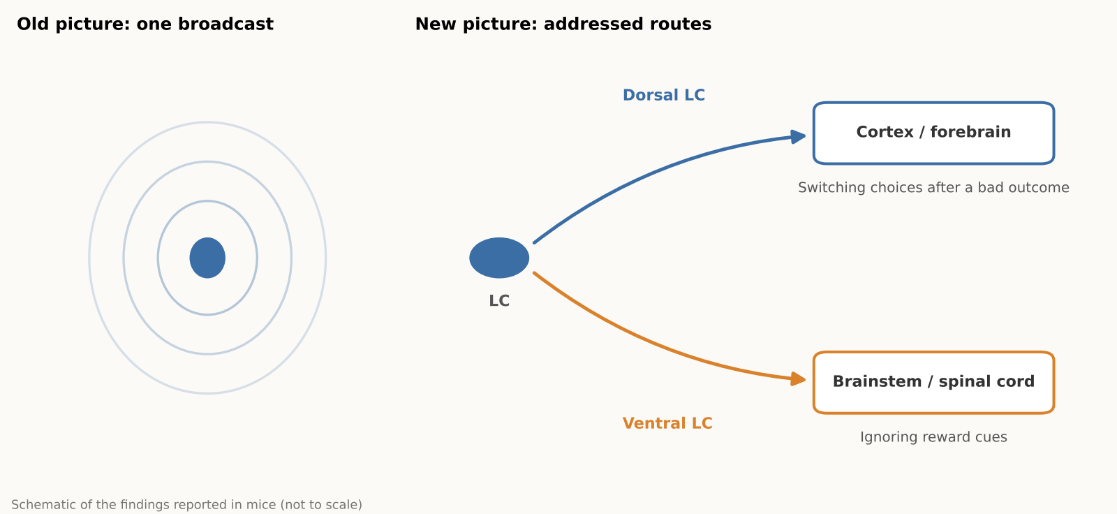
Norepinephrine, a brain chemical involved in alertness, attention and stress responses, comes largely from a tiny cluster of cells in the brainstem called the locus coeruleus (LC). For decades this system was often pictured as a foghorn, broadcasting much the same signal everywhere. A new Nature study in mice, led by researchers at the Allen Institute, finds a more organized picture, closer to a postal network: different LC neurons tend to send norepinephrine to different destinations and show different activity during behavior. Why it matters: some medications for depression and ADHD influence norepinephrine signaling, and a wiring map is a first step toward understanding how the system could one day be influenced more selectively.
Title: Topographic structure and function of locus coeruleus noradrenaline neurons
Authors: Zhixiao Su, Polina Kosillo, Kanghoon Jung et al.
Journal: Nature (2026)
DOI: 10.1038/s41586-026-11026-0
What the locus coeruleus does
The locus coeruleus (LC) is a tiny brainstem nucleus, often nicknamed the “blue spot,” and the main source of norepinephrine, also called noradrenaline, for much of the brain and spinal cord. The chemical helps regulate arousal, attention and mood, and some medications for depression and ADHD act on norepinephrine signaling. Changes in the LC have also been reported early in Alzheimer’s disease, although the timing and significance of that degeneration are still under study.
The textbook picture often treated the LC as a broadly broadcasting system, rather like a foghorn. Earlier work had already hinted at more structure. A 2020 review in Nature Reviews Neuroscience, for example, concluded that subgroups of LC neurons can encode specific cognitive processes, and a 2023 eLife study found two main electrical and structural cell types in the mouse LC. The new study maps how those differences in wiring, gene expression and activity are organized across the structure.
Mapping one tiny structure
Why map a structure this small? Because a nucleus that reaches almost the entire brain is hard to understand from the outside. Unless researchers know which neurons talk to which regions, it is difficult to say whether a drug, a stress response or a behavior is engaging the whole system or only a piece of it. Anatomy comes first, and function can be tested against it.
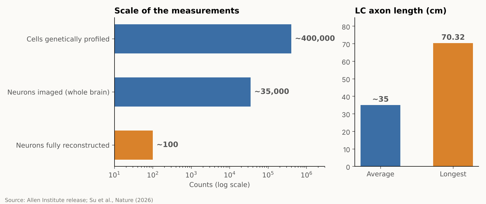
The team combined several approaches in mice, each answering a different question. Whole-brain imaging showed where LC neurons send their fibers, gene profiling showed which genes they use, and detailed reconstructions traced the branches of individual cells. The researchers then recorded neural activity while mice performed a learning task. In numbers, they imaged almost 35,000 neurons across the whole brain, profiled gene activity in nearly 400,000 cells, and fully reconstructed about 100 individual LC neurons, according to the Allen Institute. These are different measurements from different samples, not 400,000 neurons traced end to end. The research was supported by the NIH BRAIN Initiative.
A map of connections, a gradient of genes
The reconstructions showed that the LC is organized by position, as a gradient rather than two sealed compartments. Neurons in the upper (dorsal) part mainly send their axons to the cerebral cortex and forebrain, while neurons in the lower (ventral) part mainly project to the brainstem and spinal cord. Gene expression also changes smoothly along that same dorsal-to-ventral axis, matching the projection pattern, according to a summary of the paper’s abstract.
The striking part is that these patterns line up. A neuron’s position in the LC helps predict where its axon projects and how its gene expression differs from its neighbors, and, in some cases, how it behaves during learning.
A very long cell, and a selective one
LC axons, the long fibers that carry signals out of the cell, averaged about 35 centimeters in total length in the mice. The longest one traced measured 70.32 centimeters in total, which the Allen Institute describes as the longest single neuron measured in a mouse. That neuron supplies norepinephrine to a large volume of cortex but does not release it in the cerebellum, brainstem or spinal cord.
“The findings suggest the brain’s norepinephrine system is far more like a targeted postal network than a foghorn,” said Karel Svoboda of the Allen Institute.
For readers who follow how the brain learns, this fits a broader theme: learning is not one switch but many coordinated signals. We covered one such idea in our earlier piece on an MIT hypothesis about how the brain might build its own learning circuits.
Different routes, different jobs
The activity recordings pointed to different roles. Cortex-projecting dorsal neurons became more active during learning, specifically when mice changed their choice after an unfavorable outcome, and their activity also tracked reward prediction error, the gap between the reward an animal expected and what it actually got, which can guide future choices. Ventral neurons had higher background activity when mice ignored cues indicating that a reward might be available. The authors suggest this may reflect differences in how animals respond to potentially relevant cues.
A signal that carries that gap is a natural teaching tool. Finding one in a norepinephrine pathway, rather than only in the better-known dopamine system, is why the authors are interested in how the two might work together. They suspect norepinephrine and dopamine may supply complementary learning signals, but that remains a hypothesis. According to The Transmitter, the team plans to study how these chemicals interact next.
Researchers who were not involved in the work have commented on an earlier preprint version of the study. Nelson Totah of the University of Helsinki called it “bread-and-butter” research for the field that answers basic questions about an evolutionarily ancient structure. Rebecca Jordan of the University of Edinburgh said it suggests the LC could send tailored information to different brain areas, and Robert Froemke of NYU noted that the gene-expression profiling could help researchers record and manipulate specific cell types, according to The Transmitter. The published Nature paper was not evaluated by these experts in that coverage.
An independent 2025 review adds some context. It summarizes rodent studies in which ventral LC neurons reduced pain through projections to the spinal cord, while activating the LC–prefrontal circuit increased pain and anxiety-like behavior. These results come from separate experiments, but they support the idea that LC pathways can have different effects depending on their destinations.
Why it matters, and what to keep in mind
Because norepinephrine-related drugs usually act across the whole brain, a map of separate pathways raises the possibility of more selective ways to influence them that avoid unrelated circuits. The Allen Institute describes this as a possibility for the future, not a demonstrated result. All of the work was done in mice, and nobody has yet shown that human LC neurons are organized the same way. Neither does the study show that any of these pathways can be treated on its own. For now, it is a map, and a good one, rather than a medical advance.
The practical takeaway for a general reader is modest but real: the brain’s alertness and attention system is more finely organized than the old “one signal for everyone” picture suggested. That kind of basic knowledge tends to pay off slowly, by giving later researchers better targets and better questions.
Xsciens Editorial Note
The study suggests that a brain system once treated as a single volume knob is better understood as a network with different destinations. If that view is confirmed in people, treatment for focus and mood, learning support and early detection of brain aging could shift from adjusting the whole brain to targeting only the wires that matter, which may shape the precision of future drugs and diagnostics.
- What is solid is the map; what is still a hypothesis is the job of each route. Where the axons go, how gene use shifts along the structure, and the recorded activity are direct measurements, so any past interpretation that assumed one broadcast signal now needs revisiting. Reading the dorsal neurons as a “learning signal,” or the ventral ones as tied to responses to reward cues, is an interpretation drawn from how activity lines up with behavior, and the norepinephrine–dopamine partnership remains a hypothesis. These are the claims coming experiments will test.
- A mouse map is a blueprint; confirming it in people is the next stage. About 100 neurons were reconstructed in full, according to the Allen Institute, and the 35,000 imaged and 400,000 profiled cells are separate measurements. An independent 2025 review also notes that much of the evidence comes from rodents. The flip side is useful: the map tells researchers exactly what to look for in human tissue, which is where the field has to go.
- Three kinds of study to expect next (our projections). (a) Causal experiments that silence or activate one pathway at a time and see whether behavior changes, a question the study leaves open. (b) Simultaneous recordings of norepinephrine and dopamine to test whether they divide the work of learning, which The Transmitter reports the team plans to study. (c) Studies in human brains, such as post-mortem tissue or imaging, that look for the same dorsal–ventral arrangement.
- From the editors — the idea of raising or lowering a brain chemical across the board may give way to tuning one wire at a time. This map points to three futures.
1) Drug design. Today’s norepinephrine-related drugs move the pathway that serves focus and the pathway that drives bodily arousal together. The finding that gene use shifts gradually from dorsal to ventral gives researchers a possible marker for telling routes apart, and an outside expert quoted by The Transmitter noted that gene profiling could help record and manipulate specific cell types. A treatment that nudges the focus-related part without the racing heart and tension is a design goal this map makes more realistic.
2) Understanding learning. Dorsal neurons responded when mice switched choices and when outcomes missed expectations, which suggests this system may help teach us at the moment we recover from a mistake. If so, the best timing of feedback could become a design question for teaching and rehabilitation. For AI research, the idea of separating a “learn the value” signal from a “change your approach” signal into different wires may be worth borrowing.
3) Spotting change early. Changes in the LC have been reported early in Alzheimer’s disease. With this map as a ruler, researchers could ask which pathways are damaged first and whether those changes can be caught earlier and in finer detail. This study did not test that.
These are our projections, and the road from a mouse map to people is still ahead. One way to read the news in the meantime: when you see a headline about a drug that targets a particular brain pathway, check which pathway was tested in humans. That is where a map turns into a treatment, or does not.
References for this note: McKinney et al., eLife (2023), Hao et al., Int. J. Mol. Sci. (2025), The Transmitter (2026), Poe et al., Nature Reviews Neuroscience (2020)
Sources: Allen Institute, Su et al., Nature (2026), ScienceDaily