Every mammal begins life as a single fertilized egg. Through repeated divisions and a long sequence of developmental decisions, that one cell gives rise to the many specialized cells that build an animal’s body. Reconstructing exactly how those cells are related to one another has been a major scientific challenge — researchers have never had a detailed, verified map of which cell gave rise to which inside a living mammal. Two research teams have now taken a significant step toward solving that problem, publishing independently in the same week. Using DNA-based molecular recorders built into mouse embryos, they worked backward from genetic marks left behind in cells to reconstruct how a single fertilized egg becomes a full body. The work could eventually help researchers investigate how birth defects, neurodevelopmental conditions, and cancer arise.
A Lineage Record Written Into DNA
The core tool, called DNA Typewriter, was developed by a University of Washington (UW) School of Medicine team that includes geneticist Jay Shendure and researcher Junhong Choi. It is built on prime editing, a precise form of CRISPR-based gene editing that rewrites short stretches of DNA without fully cutting both strands. The team engineered a short, designated sequence into cells’ genomes and set it up so that each cell division adds one new mark, in strict order, to that sequence. Because the marks accumulate like letters typed one after another, researchers can later read the resulting sequence in any surviving cell and reconstruct which cells share a recent common ancestor and which branched apart earlier — in effect, rebuilding a family tree after the fact, rather than filming it as it happens.
Older lineage-tracing systems generally relied on CRISPR methods that cut DNA to create heritable barcodes. Those barcodes can be powerful, but as edits pile up they can become harder to read unambiguously, and some lineage information is inevitably lost along the way. Because DNA Typewriter writes new sequence without the same kind of DNA break, the team reports it can keep recording more steadily, at higher resolution, for longer than earlier cut-based approaches — though, like any method, its performance still depends on how efficiently it operates inside developing cells.
One Richly Recorded Embryo
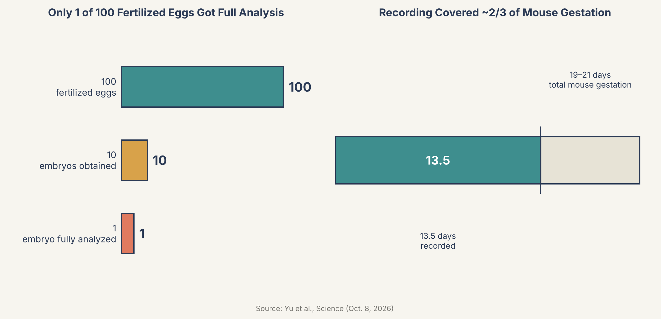
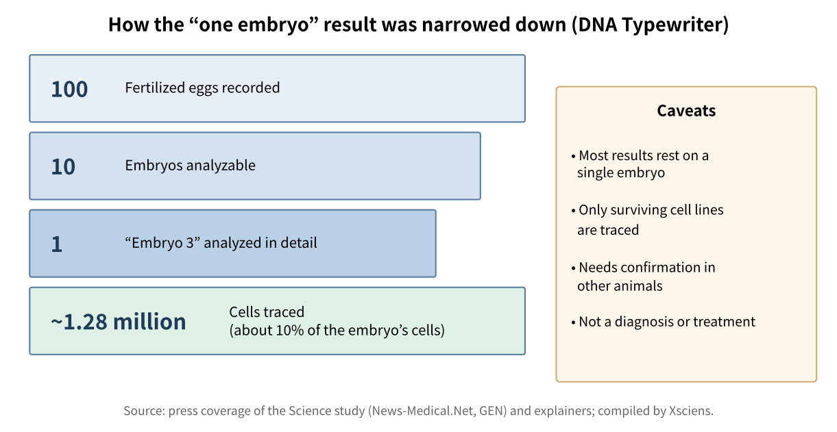
In the new study, published October 8, 2026 in the journal Science, the UW-led team — including co-first authors Qi Yu, Haedong Kim and Sophie Seidel, with Chengxiang Qiu of Dartmouth College and Shendure as co-senior authors — attempted to activate DNA Typewriter recording in 100 fertilized mouse eggs. Only 10 developed into embryos that could later be examined, and of those, one — labeled Embryo No. 3 — carried by far the richest, most complete record. That embryo became the basis for the study’s detailed lineage reconstruction, which covered roughly 1.3 million individual embryonic cells.
The recording spanned 13.5 days of development, from the fertilized egg’s first division through “late organogenesis,” the stage when the body plan and internal organs have largely taken shape. A full mouse pregnancy lasts 19 to 21 days, so the reconstruction covers a large share of gestation, though not the full life cycle. As co-first author Haedong Kim put it, the goal was a tool that “writes everything in strict order so that we can record cell lineages at much higher resolution for a longer time” than earlier barcoding methods allowed.
Two Founder Cells, One Shared Outcome
A built-in mark distinguished the two cells created when the fertilized egg first divided, letting researchers trace nearly every profiled cell in the embryo back to one of those two founders. One founder went on to produce noticeably more descendant cells than the other. Yet both founders generated a broadly similar mix of cell types in roughly equal proportions — meaning that an early imbalance in how many cells each founder produced did not translate into one founder specializing in different tissue types than the other.
The data also pointed to differences in timing: some blood and retinal cell populations showed signs of settling into a restricted identity relatively early in development, while cells contributing to the outer layer of skin appeared to keep their options open for longer. That kind of timing information — not just which cells are related, but roughly when their developmental paths narrowed — is difficult to extract from a single still snapshot of tissue, which is part of why a continuous lineage record is valuable.
A Second, Larger-Scale Study Agrees
The same week, a separate team led by Jonathan Weissman published a companion study in the journal Cell, using a related but distinct prime-editing-based system called PEtracer. Rather than following a single embryo in exhaustive detail, that study pooled data from 16 mouse embryos spanning days 7.5 to 10 of development, profiling more than 1.4 million cells in total to build a broader, population-level map of when and how cells commit to their fates. The two approaches are complementary: DNA Typewriter trades breadth for an unusually dense, ordered timeline in a single animal, while PEtracer trades timeline detail for scale across many animals. That two independently developed methods, from different labs, point toward a broadly consistent picture of mouse development adds useful weight to both — without, of course, each study individually confirming every specific claim made by the other.
“This kind of large-scale developmental recording gives scientists a map of how one cell becomes a whole body,” says Haedong Kim, co-first author of the Science study.
Weissman has framed the pair of studies as a long-sought proof of principle: that mammalian biology may finally be approaching something like the complete cell lineage map that biologist John Sulston built for the roundworm C. elegans back in 1983, by directly watching the worm’s roughly 1,000 cells divide under a microscope over years of painstaking observation. A mouse — and, by extension, a human — is built from many billions of cells, far beyond what direct visual tracking could ever handle. Genomic recording tools like DNA Typewriter and PEtracer offer a different route toward that same kind of detailed lineage map, built from molecular records and computational reconstruction rather than a person watching through a lens.
Why This Matters for Medicine
The appeal of this kind of lineage map goes beyond curiosity about development for its own sake. If researchers can see roughly how and when healthy cells commit to becoming blood, skin, or retina, they gain a reference point for recognizing what might go differently in congenital malformations, in neurodevelopmental conditions linked to early brain development, and in genetic diseases more broadly — as a way to investigate how developmental abnormalities arise, not as a diagnosis or treatment in itself. Cancer researchers are watching this space too, since tumors can, in part, involve cells that lose normal control over their fate decisions, among other changes; fine-grained lineage data is one more tool for studying how abnormal cell populations evolve over time. Xsciens has previously reported on research into how an existing cancer drug affects brain tumor cells, a reminder that untangling cellular mechanisms in detail, lineage included, is an active front across cancer research generally. Longer term, scientists studying stem cells hope lineage maps like these could help guide how lab-grown cells are coaxed into becoming the right tissue for future cell-based therapies.
It’s worth being clear about the limits here. The DNA Typewriter study’s richest results come from a single mouse embryo out of ten examined; broader validation across more animals, and eventually work beyond mice, will be needed before the tool becomes a routine research platform. Both systems have so far only been used in laboratory mouse embryos, not in humans, and translating developmental insights from mice to people remains its own multi-step scientific challenge. Even so, having two independent, broadly converging demonstrations published within the same week is a meaningful sign that genomic cell-lineage recording has moved from a promising idea toward a working research tool — one whose maps are highly informative, even if not yet complete.
Xsciens Editorial Note
“DNA wrote down how one egg becomes a body” is an appealing story, but it helps to weigh the numbers first.
- What was shown. A method that writes marks into DNA without cutting it traced a “family tree” of about 1.28 million cells over 13.5 days. A separate team’s method reported similar results, so the direction has some support.
- What is still open. The detailed analysis rests on one embryo out of 100 eggs, and the traced cells are about 10% of the embryo. Coverage of the study notes that dead cells leave no record and that confirmation in other individuals is needed.
- A caution when reading. “Clues to birth defects and cancer” in the headline is about future uses. The study itself did not demonstrate any diagnosis or treatment.
- Our imagination (speculation) — We wonder whether, as this family tree grows, disease researchers might be able to compare against a kind of standard map of “where does this cell come from when everything is normal?” That could make it easier to discuss at which branching step a congenital condition arises. But until the method works reliably across many individuals, that remains a long way off, and this is our speculation.
References for this note: The Brighter Side, “Scientists construct a ‘family tree’ containing 1.28 million cells from a single embryo” (figures from the Science study and the limitations it lists)
Sources: News-Medical.Net, Genetic Engineering & Biotechnology News (GEN)
Leave a Reply